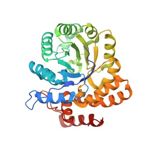
Structural and Biochemical Studies of Human 4-hydroxy-2-oxoglutarate Aldolase: Implications for Hydroxyproline Metabolism in Primary Hyperoxaluria.
Riedel, T.J., Johnson, L.C., Knight, J., Hantgan, R.R., Holmes, R.P., Lowther, W.T.(2011) PLoS One 6: e26021-e26021
- PubMed: 21998747 Search on PubMedSearch on PubMed Central
- DOI: https://doi.org/10.1371/journal.pone.0026021
- Primary Citation Related Structures:
3S5N, 3S5O - PubMed Abstract:
4-hydroxy-2-oxoglutarate (HOG) aldolase is a unique enzyme in the hydroxyproline degradation pathway catalyzing the cleavage of HOG to pyruvate and glyoxylate. Mutations in this enzyme are believed to be associated with the excessive production of oxalate in primary hyperoxaluria type 3 (PH3), although no experimental data is available to support this hypothesis. Moreover, the identity, oligomeric state, enzymatic activity, and crystal structure of human HOGA have not been experimentally determined. In this study human HOGA (hHOGA) was identified by mass spectrometry of the mitochondrial enzyme purified from bovine kidney. hHOGA performs a retro-aldol cleavage reaction reminiscent of the trimeric 2-keto-3-deoxy-6-phosphogluconate aldolases. Sequence comparisons, however, show that HOGA is related to the tetrameric, bacterial dihydrodipicolinate synthases, but the reaction direction is reversed. The 1.97 Å resolution crystal structure of hHOGA bound to pyruvate was determined and enabled the modeling of the HOG-Schiff base intermediate and the identification of active site residues. Kinetic analyses of site-directed mutants support the importance of Lys196 as the nucleophile, Tyr168 and Ser77 as components of a proton relay, and Asn78 and Ser198 as unique residues that facilitate substrate binding. The biochemical and structural data presented support that hHOGA utilizes a type I aldolase reaction mechanism, but employs novel residue interactions for substrate binding. A mapping of the PH3 mutations identifies potential rearrangements in either the active site or the tetrameric assembly that would likely cause a loss in activity. Altogether, these data establish a foundation to assess mutant forms of hHOGA and how their activity could be pharmacologically restored.
- Center for Structural Biology and Department of Biochemistry, Wake Forest School of Medicine, Winston-Salem, North Carolina, United States of America.
Organizational Affiliation: