3S48

Human Alpha-Haemoglobin Complexed with the First NEAT Domain of IsdH from Staphylococcus aureus


Experimental Data Snapshot

  • Method: X-RAY DIFFRACTION
  • Resolution: 3.05 Å
  • R-Value Free: 0.268 
  • R-Value Work: 0.240 
  • R-Value Observed: 0.242 

wwPDB Validation   3D Report Full Report


Ligand Structure Quality Assessment 


This is version 1.2 of the entry. See complete history


Literature

The structure of alpha-haemoglobin in complex with a haemoglobin-binding domain from Staphylococcus aureus reveals the elusive alpha-haemoglobin dimerization interface

Krishna Kumar, K.Jacques, D.A.Guss, J.M.Gell, D.A.

(2014) Acta Crystallogr F Struct Biol Commun 70: 1032-1037

  • DOI: https://doi.org/10.1107/S2053230X14012175
  • Primary Citation of Related Structures:  
    3S48

  • PubMed Abstract: 

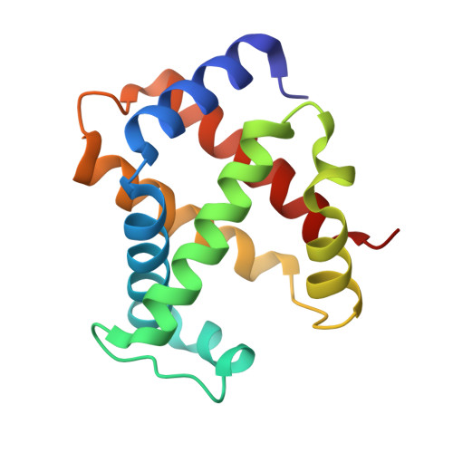
    Adult haemoglobin (Hb) is made up of two α and two β subunits. Mutations that reduce expression of the α- or β-globin genes lead to the conditions α- or β-thalassaemia, respectively. Whilst both conditions are characterized by anaemia of variable severity, other details of their pathophysiology are different, in part owing to the greater stability of the β chains that is conferred through β self-association. In contrast, α subunits interact weakly, and in the absence of stabilizing quaternary interactions the α chain (α) is prone to haem loss and denaturation. The molecular contacts that confer weak self-association of α have not been determined previously. Here, the first structure of an α2 homodimer is reported in complex with one domain of the Hb receptor from Staphylococcus aureus. The α2 dimer interface has a highly unusual, approximately linear, arrangement of four His side chains within hydrogen-bonding distance of each other. Some interactions present in the α1β1 dimer interface of native Hb are preserved in the α2 dimer. However, a marked asymmetry is observed in the α2 interface, suggesting that steric factors limit the number of stabilizing interactions that can form simultaneously across the interface.


  • Organizational Affiliation

    School of Molecular Bioscience, University of Sydney, Sydney, NSW 2006, Australia.


Macromolecules
Find similar proteins by:  (by identity cutoff)  |  3D Structure
Entity ID: 1
MoleculeChains Sequence LengthOrganismDetailsImage
Iron-regulated surface determinant protein HA,
C [auth B]
164Staphylococcus aureus subsp. aureus MSSA476Mutation(s): 0 
Gene Names: isdH
UniProt
Find proteins for Q6G8J7 (Staphylococcus aureus (strain MSSA476))
Explore Q6G8J7 
Go to UniProtKB:  Q6G8J7
Entity Groups  
Sequence Clusters30% Identity50% Identity70% Identity90% Identity95% Identity100% Identity
UniProt GroupQ6G8J7
Sequence Annotations
Expand
  • Reference Sequence
Find similar proteins by:  (by identity cutoff)  |  3D Structure
Entity ID: 2
MoleculeChains Sequence LengthOrganismDetailsImage
Hemoglobin subunit alphaB [auth D],
D [auth C]
141Homo sapiensMutation(s): 0 
UniProt & NIH Common Fund Data Resources
Find proteins for P69905 (Homo sapiens)
Explore P69905 
Go to UniProtKB:  P69905
PHAROS:  P69905
Entity Groups  
Sequence Clusters30% Identity50% Identity70% Identity90% Identity95% Identity100% Identity
UniProt GroupP69905
Sequence Annotations
Expand
  • Reference Sequence
Small Molecules
Ligands 1 Unique
IDChains Name / Formula / InChI Key2D Diagram3D Interactions
HEM
Query on HEM

Download Ideal Coordinates CCD File 
E [auth D],
F [auth C]
PROTOPORPHYRIN IX CONTAINING FE
C34 H32 Fe N4 O4
KABFMIBPWCXCRK-RGGAHWMASA-L
Experimental Data & Validation

Experimental Data

  • Method: X-RAY DIFFRACTION
  • Resolution: 3.05 Å
  • R-Value Free: 0.268 
  • R-Value Work: 0.240 
  • R-Value Observed: 0.242 
  • Space Group: I 41
Unit Cell:
Length ( Å )Angle ( ˚ )
a = 115.607α = 90
b = 115.607β = 90
c = 142.333γ = 90
Software Package:
Software NamePurpose
Blu-Icedata collection
PHASERphasing
REFMACrefinement
HKL-2000data reduction
SCALEPACKdata scaling

Structure Validation

View Full Validation Report


Ligand Structure Quality Assessment 


Entry History 

Deposition Data

Revision History  (Full details and data files)

  • Version 1.0: 2012-05-23
    Type: Initial release
  • Version 1.1: 2014-08-20
    Changes: Database references
  • Version 1.2: 2023-11-01
    Changes: Data collection, Database references, Derived calculations, Refinement description