3RYO | pdb_00003ryo

Crystal Structure of Enhanced Intracellular Survival (Eis) Protein from Mycobacterium tuberculosis with Acetyl CoA


Experimental Data Snapshot

  • Method: X-RAY DIFFRACTION
  • Resolution: 2.80 Å
  • R-Value Free: 
    0.248 (Depositor), 0.217 (DCC) 
  • R-Value Work: 
    0.192 (Depositor), 0.219 (DCC) 
  • R-Value Observed: 
    0.195 (Depositor) 

wwPDB Validation 3D Report Full Report

Validation slider image for 3RYO

Ligand Structure Quality Assessment 


This is version 1.1 of the entry. See complete history

Literature

Mycobacterium tuberculosis Eis protein initiates suppression of host immune responses by acetylation of DUSP16/MKP-7

Kim, K.H.An, D.R.Song, J.Yoon, J.Y.Kim, H.S.Yoon, H.J.Im, H.N.Kim, J.Kim, D.J.Lee, S.J.Kim, K.H.Lee, H.M.Kim, H.J.Jo, E.K.Lee, J.Y.Suh, S.W.

(2012) Proc Natl Acad Sci U S A 

  • DOI: https://doi.org/10.1073/pnas.1120251109
  • Primary Citation Related Structures: 
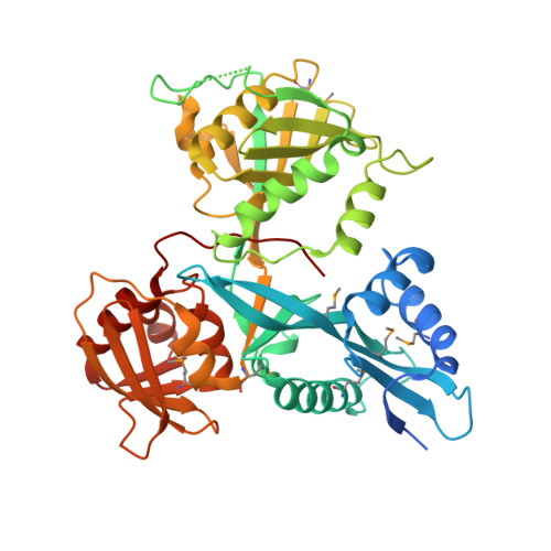
    3RYO, 3SXN, 3UY5

  • PubMed Abstract: 

    The intracellular pathogen Mycobacterium tuberculosis (Mtb) causes tuberculosis. Enhanced intracellular survival (Eis) protein, secreted by Mtb, enhances survival of Mycobacterium smegmatis (Msm) in macrophages. Mtb Eis was shown to suppress host immune defenses by negatively modulating autophagy, inflammation, and cell death through JNK-dependent inhibition of reactive oxygen species (ROS) generation. Mtb Eis was recently demonstrated to contribute to drug resistance by acetylating multiple amines of aminoglycosides. However, the mechanism of enhanced intracellular survival by Mtb Eis remains unanswered. Therefore, we have characterized both Mtb and Msm Eis proteins biochemically and structurally. We have discovered that Mtb Eis is an efficient N(ε)-acetyltransferase, rapidly acetylating Lys55 of dual-specificity protein phosphatase 16 (DUSP16)/mitogen-activated protein kinase phosphatase-7 (MKP-7), a JNK-specific phosphatase. In contrast, Msm Eis is more efficient as an N(α)-acetyltransferase. We also show that Msm Eis acetylates aminoglycosides as readily as Mtb Eis. Furthermore, Mtb Eis, but not Msm Eis, inhibits LPS-induced JNK phosphorylation. This functional difference against DUSP16/MKP-7 can be understood by comparing the structures of two Eis proteins. The active site of Mtb Eis with a narrow channel seems more suitable for sequence-specific recognition of the protein substrate than the pocket-shaped active site of Msm Eis. We propose that Mtb Eis initiates the inhibition of JNK-dependent autophagy, phagosome maturation, and ROS generation by acetylating DUSP16/MKP-7. Our work thus provides insight into the mechanism of suppressing host immune responses and enhancing mycobacterial survival within macrophages by Mtb Eis.


  • Organizational Affiliation
    • Department of Chemistry, College of Natural Sciences, Seoul National University, Seoul 151-742, Korea.

Macromolecule Content 

  • Total Structure Weight: 574.52 kDa 
  • Atom Count: 37,731 
  • Modeled Residue Count: 4,752 
  • Deposited Residue Count: 5,136 
  • Unique protein chains: 1

Macromolecules

Find similar proteins by:|  3D Structure
Entity ID: 1
MoleculeChains  Sequence LengthOrganismDetailsImage
Enhanced intracellular survival protein
A, B, C, D, E
A, B, C, D, E, F, G, H, I, J, K, L
428Mycobacterium tuberculosisMutation(s): 0 
Gene Names: eisRv2416cMT2489MTCY253.04
EC: 2.3.1
UniProt
Find proteins for P9WFK7 (Mycobacterium tuberculosis (strain ATCC 25618 / H37Rv))
Explore P9WFK7 
Go to UniProtKB:  P9WFK7
Entity Groups
Sequence Clusters30% Identity50% Identity70% Identity90% Identity95% Identity100% Identity
UniProt GroupP9WFK7
Sequence Annotations
Expand
Reference Sequence

Small Molecules

Ligands 1 Unique
IDChains Name / Formula / InChI Key2D Diagram3D Interactions
ACO

Query on ACO


Download:Ideal Coordinates CCD File
M [auth A]
N [auth B]
O [auth C]
P [auth D]
Q [auth E]
M [auth A],
N [auth B],
O [auth C],
P [auth D],
Q [auth E],
R [auth F],
S [auth G],
T [auth H],
U [auth I],
V [auth J],
W [auth K],
X [auth L]
ACETYL COENZYME *A
C23 H38 N7 O17 P3 S
ZSLZBFCDCINBPY-ZSJPKINUSA-N
Modified Residues  1 Unique
IDChains TypeFormula2D DiagramParent
MSE
Query on MSE
A, B, C, D, E
A, B, C, D, E, F, G, H, I, J, K, L
L-PEPTIDE LINKINGC5 H11 N O2 SeMET

Experimental Data & Validation

Experimental Data

  • Method: X-RAY DIFFRACTION
  • Resolution: 2.80 Å
  • R-Value Free:  0.248 (Depositor), 0.217 (DCC) 
  • R-Value Work:  0.192 (Depositor), 0.219 (DCC) 
  • R-Value Observed: 0.195 (Depositor) 
Space Group: P 1 21 1
Unit Cell:
Length ( Å )Angle ( ˚ )
a = 108.129α = 90
b = 150.19β = 103.05
c = 184.383γ = 90
Software Package:
Software NamePurpose
PHENIXrefinement
PDB_EXTRACTdata extraction

Structure Validation

View Full Validation Report


Ligand Structure Quality Assessment 


Entry History 

Deposition Data

Revision History  (Full details and data files)

  • Version 1.0: 2012-05-23
    Type: Initial release
  • Version 1.1: 2024-10-09
    Changes: Data collection, Database references, Derived calculations, Structure summary