3RYL | pdb_00003ryl

Dimerization domain of Vibrio parahemolyticus VopL


Experimental Data Snapshot

  • Method: X-RAY DIFFRACTION
  • Resolution: 3.10 Å
  • R-Value Free: 
    0.294 (Depositor), 0.288 (DCC) 
  • R-Value Work: 
    0.240 (Depositor), 0.232 (DCC) 
  • R-Value Observed: 
    0.246 (Depositor) 

wwPDB Validation 3D Report Full Report

Validation slider image for 3RYL

This is version 1.2 of the entry. See complete history

Literature

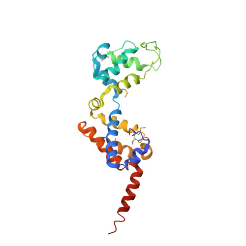
Mechanism of actin filament nucleation by Vibrio VopL and implications for tandem W domain nucleation.

Namgoong, S.Boczkowska, M.Glista, M.J.Winkelman, J.D.Rebowski, G.Kovar, D.R.Dominguez, R.

(2011) Nat Struct Mol Biol 18: 1060-1067

  • DOI: https://doi.org/10.1038/nsmb.2109
  • Primary Citation Related Structures: 
    3RYL

  • PubMed Abstract: 

    Pathogen proteins targeting the actin cytoskeleton often serve as model systems to understand their more complex eukaryotic analogs. We show that the strong actin filament nucleation activity of Vibrio parahaemolyticus VopL depends on its three W domains and on its dimerization through a unique VopL C-terminal domain (VCD). The VCD shows a previously unknown all-helical fold and interacts with the pointed end of the actin nucleus, contributing to the nucleation activity directly and through duplication of the W domain repeat. VopL promotes rapid cycles of filament nucleation and detachment but generally has no effect on elongation. Profilin inhibits VopL-induced nucleation by competing for actin binding to the W domains. Combined, the results suggest that VopL stabilizes a hexameric double-stranded pointed end nucleus. Analysis of hybrid constructs of VopL and the eukaryotic nucleator Spire suggest that Spire may also function as a dimer in cells.


  • Organizational Affiliation
    • Department of Physiology, Perelman School of Medicine, University of Pennsylvania, Philadelphia, Pennsylvania, USA.

Macromolecule Content 

  • Total Structure Weight: 54.13 kDa 
  • Atom Count: 3,484 
  • Modeled Residue Count: 440 
  • Deposited Residue Count: 482 
  • Unique protein chains: 1

Macromolecules

Find similar proteins by:|  3D Structure
Entity ID: 1
MoleculeChains  Sequence LengthOrganismDetailsImage
protein VPA1370
A, B
241Vibrio parahaemolyticus RIMD 2210633Mutation(s): 0 
Gene Names: VopLVPA1370
UniProt
Find proteins for Q87GE5 (Vibrio parahaemolyticus serotype O3:K6 (strain RIMD 2210633))
Explore Q87GE5 
Go to UniProtKB:  Q87GE5
Entity Groups
Sequence Clusters30% Identity50% Identity70% Identity90% Identity95% Identity100% Identity
UniProt GroupQ87GE5
Sequence Annotations
Expand
Reference Sequence

Small Molecules

Modified Residues  1 Unique
IDChains TypeFormula2D DiagramParent
MSE
Query on MSE
A, B
L-PEPTIDE LINKINGC5 H11 N O2 SeMET

Experimental Data & Validation

Experimental Data

  • Method: X-RAY DIFFRACTION
  • Resolution: 3.10 Å
  • R-Value Free:  0.294 (Depositor), 0.288 (DCC) 
  • R-Value Work:  0.240 (Depositor), 0.232 (DCC) 
  • R-Value Observed: 0.246 (Depositor) 
Space Group: P 21 21 21
Unit Cell:
Length ( Å )Angle ( ˚ )
a = 56.93α = 90
b = 90.49β = 90
c = 101.86γ = 90
Software Package:
Software NamePurpose
ADSCdata collection
SHELXSphasing
PHENIXrefinement
XDSdata reduction
XSCALEdata scaling

Structure Validation

View Full Validation Report


Entry History 

Deposition Data

Revision History  (Full details and data files)

  • Version 1.0: 2011-08-31
    Type: Initial release
  • Version 1.1: 2013-06-19
    Changes: Database references
  • Version 1.2: 2024-10-30
    Changes: Data collection, Database references, Derived calculations, Structure summary