3RO9

Candida glabrata dihydrofolate reductase complexed with NADPH and 6-ethyl-5-[(3R)-3-[3-methoxy-5-(pyridin-4-yl)phenyl]but-1-yn-1-yl]pyrimidine-2,4-diamine (UCP1006)


Experimental Data Snapshot

  • Method: X-RAY DIFFRACTION
  • Resolution: 2.60 Å
  • R-Value Free: 0.265 
  • R-Value Work: 0.202 
  • R-Value Observed: 0.205 

wwPDB Validation   3D Report Full Report


Ligand Structure Quality Assessment 


This is version 1.4 of the entry. See complete history


Literature

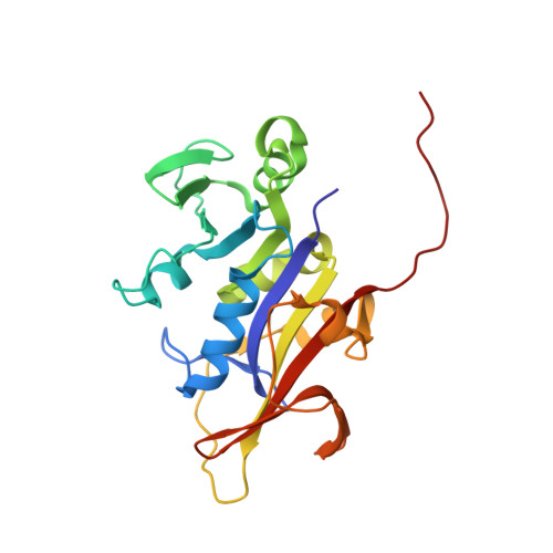
Structural analysis of the active sites of dihydrofolate reductase from two species of Candida uncovers ligand-induced conformational changes shared among species.

Paulsen, J.L.Viswanathan, K.Wright, D.L.Anderson, A.C.

(2013) Bioorg Med Chem Lett 23: 1279-1284

  • DOI: https://doi.org/10.1016/j.bmcl.2013.01.008
  • Primary Citation of Related Structures:  
    3RO9, 3ROA, 4H95, 4H96, 4H97, 4H98

  • PubMed Abstract: 

    A novel strategy for targeting the pathogenic organisms Candida albicans and Candida glabrata focuses on the development of potent and selective antifolates effective against dihydrofolate reductase. Crystal structure analysis suggested that an essential loop at the active site (Thr 58-Phe 66) differs from the analogous residues in the human enzyme, potentially providing a mechanism for achieving selectivity. In order to probe the role of this loop, we employed chemical synthesis, crystal structure determination and molecular dynamics simulations. The results of these analyses show that the loop residues undergo ligand-induced conformational changes that are similar among the fungal and human species.


  • Organizational Affiliation

    Department of Pharmaceutical Sciences, University of Connecticut, 69 N. Eagleville Rd., Storrs, CT 06269, USA.


Macromolecules
Find similar proteins by:  (by identity cutoff)  |  3D Structure
Entity ID: 1
MoleculeChains Sequence LengthOrganismDetailsImage
Strain CBS138 chromosome J complete sequence
A, B
225Nakaseomyces glabratusMutation(s): 0 
Gene Names: CAGL0J03894gDHFR
UniProt
Find proteins for Q6FPH0 (Candida glabrata (strain ATCC 2001 / BCRC 20586 / JCM 3761 / NBRC 0622 / NRRL Y-65 / CBS 138))
Explore Q6FPH0 
Go to UniProtKB:  Q6FPH0
Entity Groups  
Sequence Clusters30% Identity50% Identity70% Identity90% Identity95% Identity100% Identity
UniProt GroupQ6FPH0
Sequence Annotations
Expand
  • Reference Sequence
Small Molecules
Ligands 2 Unique
IDChains Name / Formula / InChI Key2D Diagram3D Interactions
NDP
Query on NDP

Download Ideal Coordinates CCD File 
C [auth A],
E [auth B]
NADPH DIHYDRO-NICOTINAMIDE-ADENINE-DINUCLEOTIDE PHOSPHATE
C21 H30 N7 O17 P3
ACFIXJIJDZMPPO-NNYOXOHSSA-N
06U
Query on 06U

Download Ideal Coordinates CCD File 
D [auth A],
F [auth B]
6-ethyl-5-{(3R)-3-[3-methoxy-5-(pyridin-4-yl)phenyl]but-1-yn-1-yl}pyrimidine-2,4-diamine
C22 H23 N5 O
KEPLBUUTAQCZOE-AWEZNQCLSA-N
Experimental Data & Validation

Experimental Data

  • Method: X-RAY DIFFRACTION
  • Resolution: 2.60 Å
  • R-Value Free: 0.265 
  • R-Value Work: 0.202 
  • R-Value Observed: 0.205 
  • Space Group: P 41
Unit Cell:
Length ( Å )Angle ( ˚ )
a = 42.26α = 90
b = 42.26β = 90
c = 238.69γ = 90
Software Package:
Software NamePurpose
SCALEPACKdata scaling
PHASERphasing
REFMACrefinement
PDB_EXTRACTdata extraction
HKL-2000data collection
DENZOdata reduction

Structure Validation

View Full Validation Report


Ligand Structure Quality Assessment 


Entry History 

Deposition Data

Revision History  (Full details and data files)

  • Version 1.0: 2012-05-09
    Type: Initial release
  • Version 1.1: 2013-03-27
    Changes: Database references
  • Version 1.2: 2013-11-20
    Changes: Non-polymer description
  • Version 1.3: 2017-11-08
    Changes: Refinement description
  • Version 1.4: 2024-02-28
    Changes: Data collection, Database references, Derived calculations, Refinement description