3RN5 | pdb_00003rn5

Structural basis of cytosolic DNA recognition by innate immune receptors


Experimental Data Snapshot

  • Method: X-RAY DIFFRACTION
  • Resolution: 2.50 Å
  • R-Value Free: 
    0.241 (Depositor), 0.243 (DCC) 
  • R-Value Work: 
    0.201 (Depositor), 0.201 (DCC) 
  • R-Value Observed: 
    0.202 (Depositor) 

Starting Model: experimental
View more details

wwPDB Validation 3D Report Full Report

Validation slider image for 3RN5

This is version 1.2 of the entry. See complete history

Literature

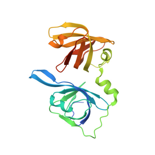
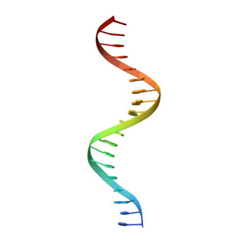
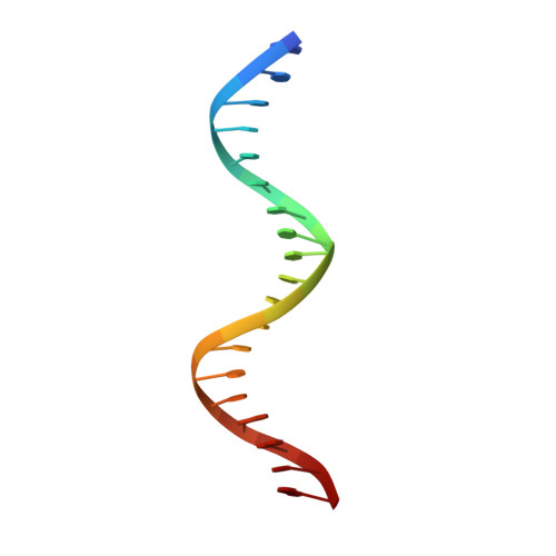
Structures of the HIN Domain:DNA Complexes Reveal Ligand Binding and Activation Mechanisms of the AIM2 Inflammasome and IFI16 Receptor.

Jin, T.Perry, A.Jiang, J.Smith, P.Curry, J.A.Unterholzner, L.Jiang, Z.Horvath, G.Rathinam, V.A.Johnstone, R.W.Hornung, V.Latz, E.Bowie, A.G.Fitzgerald, K.A.Xiao, T.S.

(2012) Immunity 36: 561-571

  • DOI: https://doi.org/10.1016/j.immuni.2012.02.014
  • Primary Citation Related Structures: 
    3RLN, 3RLO, 3RN2, 3RN5, 3RNU

  • PubMed Abstract: 

    Recognition of DNA by the innate immune system is central to antiviral and antibacterial defenses, as well as an important contributor to autoimmune diseases involving self DNA. AIM2 (absent in melanoma 2) and IFI16 (interferon-inducible protein 16) have been identified as DNA receptors that induce inflammasome formation and interferon production, respectively. Here we present the crystal structures of their HIN domains in complex with double-stranded (ds) DNA. Non-sequence-specific DNA recognition is accomplished through electrostatic attraction between the positively charged HIN domain residues and the dsDNA sugar-phosphate backbone. An intramolecular complex of the AIM2 Pyrin and HIN domains in an autoinhibited state is liberated by DNA binding, which may facilitate the assembly of inflammasomes along the DNA staircase. These findings provide mechanistic insights into dsDNA as the activation trigger and oligomerization platform for the assembly of large innate signaling complexes such as the inflammasomes.


  • Organizational Affiliation
    • Structural Immunobiology Unit, Laboratory of Immunology, National Institute of Allergy and Infectious Diseases, National Institutes of Health, Bethesda, MD 20892-0430, USA.

Macromolecule Content 

  • Total Structure Weight: 117.78 kDa 
  • Atom Count: 7,944 
  • Modeled Residue Count: 851 
  • Deposited Residue Count: 908 
  • Unique protein chains: 1
  • Unique nucleic acid chains: 2

Macromolecules


Find similar proteins by:|  3D Structure
Entity ID: 1
MoleculeChains  Sequence LengthOrganismDetailsImage
Interferon-inducible protein AIM2
A, B, C, D
208Homo sapiensMutation(s): 0 
Gene Names: AIM2
UniProt & NIH Common Fund Data Resources
Find proteins for O14862 (Homo sapiens)
Explore O14862 
Go to UniProtKB:  O14862
PHAROS:  O14862
GTEx:  ENSG00000163568 
Entity Groups
Sequence Clusters30% Identity50% Identity70% Identity90% Identity95% Identity100% Identity
UniProt GroupO14862
Sequence Annotations
Expand
Reference Sequence
Find similar nucleic acids by:  Sequence
Entity ID: 2
MoleculeChains LengthOrganismImage
DNA (5'-D(*CP*CP*AP*TP*CP*AP*AP*AP*GP*AP*GP*AP*GP*AP*AP*AP*GP*AP*G)-3')E [auth K],
G [auth M]
19N/A
Sequence Annotations
Expand
Reference Sequence
Find similar nucleic acids by:  Sequence
Entity ID: 3
MoleculeChains LengthOrganismImage
DNA (5'-D(*GP*CP*TP*CP*TP*TP*TP*CP*TP*CP*TP*CP*TP*TP*TP*GP*AP*TP*G)-3')F [auth L],
H [auth N]
19N/A
Sequence Annotations
Expand
Reference Sequence

Small Molecules

Ligands 1 Unique
IDChains Name / Formula / InChI Key2D Diagram3D Interactions
EDO

Query on EDO


Download:Ideal Coordinates CCD File
I [auth A]
J [auth A]
K [auth B]
L [auth B]
M [auth C]
I [auth A],
J [auth A],
K [auth B],
L [auth B],
M [auth C],
N [auth D],
O [auth D],
P [auth D],
Q [auth D]
1,2-ETHANEDIOL
C2 H6 O2
LYCAIKOWRPUZTN-UHFFFAOYSA-N

Experimental Data & Validation

Experimental Data

  • Method: X-RAY DIFFRACTION
  • Resolution: 2.50 Å
  • R-Value Free:  0.241 (Depositor), 0.243 (DCC) 
  • R-Value Work:  0.201 (Depositor), 0.201 (DCC) 
  • R-Value Observed: 0.202 (Depositor) 
Space Group: P 1 21 1
Unit Cell:
Length ( Å )Angle ( ˚ )
a = 62.36α = 90
b = 136.36β = 89.98
c = 77.04γ = 90
Software Package:
Software NamePurpose
Blu-Icedata collection
PHASERphasing
PHENIXrefinement
XDSdata reduction
XDSdata scaling

Structure Validation

View Full Validation Report


Entry History 

Deposition Data

Revision History  (Full details and data files)

  • Version 1.0: 2012-04-25
    Type: Initial release
  • Version 1.1: 2012-05-09
    Changes: Database references
  • Version 1.2: 2023-09-13
    Changes: Data collection, Database references, Derived calculations, Refinement description