Identification of the bile salt binding site on IpaD from Shigella flexneri and the influence of ligand binding on IpaD structure.
Barta, M.L., Guragain, M., Adam, P., Dickenson, N.E., Patil, M., Geisbrecht, B.V., Picking, W.L., Picking, W.D.(2012) Proteins 80: 935-945
- PubMed: 22423359 Search on PubMedSearch on PubMed Central
- DOI: https://doi.org/10.1002/prot.23251
- Primary Citation Related Structures:
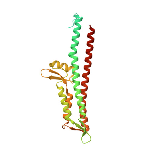
3R9V - PubMed Abstract:
Type III secretion (TTS) is an essential virulence factor for Shigella flexneri, the causative agent of shigellosis. The Shigella TTS apparatus (TTSA) is an elegant nanomachine that is composed of a basal body, an external needle to deliver effectors into human cells, and a needle tip complex that controls secretion activation. IpaD is at the tip of the nascent TTSA needle where it controls the first step of TTS activation. The bile salt deoxycholate (DOC) binds to IpaD to induce recruitment of the translocator protein IpaB into the maturing tip complex. We recently used spectroscopic analyses to show that IpaD undergoes a structural rearrangement that accompanies binding to DOC. Here, we report a crystal structure of IpaD with DOC bound and test the importance of the residues that make up the DOC binding pocket on IpaD function. IpaD binds DOC at the interface between helices α3 and α7, with concomitant movement in the orientation of helix α7 relative to its position in unbound IpaD. When the IpaD residues involved in DOC binding are mutated, some are found to lead to altered invasion and secretion phenotypes. These findings suggest that adoption of a DOC bound structural state for IpaD primes the Shigella TTSA for contact with host cells. The data presented here and in the studies leading up to this work provide the foundation for developing a model of the first step in Shigella TTS activation.
- Division of Cell Biology, School of Biological Sciences, University of Missouri-Kansas City, Kansas City, Missouri, USA.
Organizational Affiliation: