3R5W | pdb_00003r5w

Structure of Ddn, the Deazaflavin-dependent nitroreductase from Mycobacterium tuberculosis involved in bioreductive activation of PA-824, with co-factor F420


Experimental Data Snapshot

  • Method: X-RAY DIFFRACTION
  • Resolution: 1.79 Å
  • R-Value Free: 
    0.206 (Depositor), 0.211 (DCC) 
  • R-Value Work: 
    0.173 (Depositor), 0.179 (DCC) 
  • R-Value Observed: 
    0.175 (Depositor) 

Starting Model: experimental
View more details

wwPDB Validation 3D Report Full Report

Validation slider image for 3R5W

Ligand Structure Quality Assessment 


This is version 1.3 of the entry. See complete history

Literature

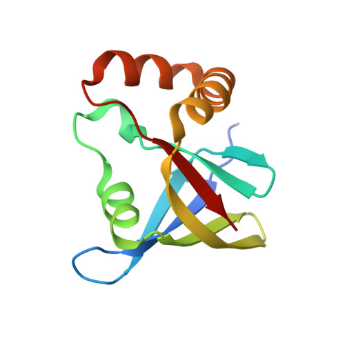
Structure of Ddn, the deazaflavin-dependent nitroreductase from Mycobacterium tuberculosis involved in bioreductive activation of PA-824.

Cellitti, S.E.Shaffer, J.Jones, D.H.Mukherjee, T.Gurumurthy, M.Bursulaya, B.Boshoff, H.I.Choi, I.Nayyar, A.Lee, Y.S.Cherian, J.Niyomrattanakit, P.Dick, T.Manjunatha, U.H.Barry, C.E.Spraggon, G.Geierstanger, B.H.

(2012) Structure 20: 101-112

  • DOI: https://doi.org/10.1016/j.str.2011.11.001
  • Primary Citation Related Structures: 
    3R5L, 3R5P, 3R5R, 3R5W, 3R5Y, 3R5Z

  • PubMed Abstract: 

    Tuberculosis continues to be a global health threat, making bicyclic nitroimidazoles an important new class of therapeutics. A deazaflavin-dependent nitroreductase (Ddn) from Mycobacterium tuberculosis catalyzes the reduction of nitroimidazoles such as PA-824, resulting in intracellular release of lethal reactive nitrogen species. The N-terminal 30 residues of Ddn are functionally important but are flexible or access multiple conformations, preventing structural characterization of the full-length, enzymatically active enzyme. Several structures were determined of a truncated, inactive Ddn protein core with and without bound F(420) deazaflavin coenzyme as well as of a catalytically competent homolog from Nocardia farcinica. Mutagenesis studies based on these structures identified residues important for binding of F(420) and PA-824. The proposed orientation of the tail of PA-824 toward the N terminus of Ddn is consistent with current structure-activity relationship data.


  • Organizational Affiliation
    • Genomics Institute of the Novartis Research Foundation, 10675 John Jay Hopkins Drive, San Diego, CA 92121-1125, USA.

Macromolecule Content 

  • Total Structure Weight: 135.3 kDa 
  • Atom Count: 10,600 
  • Modeled Residue Count: 1,090 
  • Deposited Residue Count: 1,120 
  • Unique protein chains: 1

Macromolecules

Find similar proteins by:|  3D Structure
Entity ID: 1
MoleculeChains  Sequence LengthOrganismDetailsImage
Deazaflavin-dependent nitroreductase112Mycobacterium tuberculosisMutation(s): 0 
Gene Names: ddnMT3651Rv3547
EC: 1 (PDB Primary Data), 1.1.98 (UniProt)
UniProt
Find proteins for P9WP15 (Mycobacterium tuberculosis (strain ATCC 25618 / H37Rv))
Explore P9WP15 
Go to UniProtKB:  P9WP15
Entity Groups
Sequence Clusters30% Identity50% Identity70% Identity90% Identity95% Identity100% Identity
UniProt GroupP9WP15
Sequence Annotations
Expand
Reference Sequence

Small Molecules

Ligands 1 Unique
IDChains Name / Formula / InChI Key2D Diagram3D Interactions
F42

Query on F42


Download:Ideal Coordinates CCD File
K [auth A]
L [auth B]
M [auth C]
N [auth D]
O [auth E]
K [auth A],
L [auth B],
M [auth C],
N [auth D],
O [auth E],
P [auth K],
Q [auth L],
R [auth M],
S [auth N],
T [auth O]
COENZYME F420
C29 H36 N5 O18 P
GEHSZWRGPHDXJO-NALJQGANSA-N

Experimental Data & Validation

Experimental Data

  • Method: X-RAY DIFFRACTION
  • Resolution: 1.79 Å
  • R-Value Free:  0.206 (Depositor), 0.211 (DCC) 
  • R-Value Work:  0.173 (Depositor), 0.179 (DCC) 
  • R-Value Observed: 0.175 (Depositor) 
Space Group: P 1 21 1
Unit Cell:
Length ( Å )Angle ( ˚ )
a = 85.336α = 90
b = 89.419β = 96.29
c = 127.678γ = 90
Software Package:
Software NamePurpose
DENZOdata reduction
SCALEPACKdata scaling
PHASERphasing
PHENIXrefinement
PDB_EXTRACTdata extraction
BOSdata collection
HKL-2000data reduction
HKL-2000data scaling

Structure Validation

View Full Validation Report


Ligand Structure Quality Assessment 


Entry History 

Revision History  (Full details and data files)

  • Version 1.0: 2012-01-18
    Type: Initial release
  • Version 1.1: 2012-03-21
    Changes: Database references
  • Version 1.2: 2017-11-08
    Changes: Refinement description
  • Version 1.3: 2023-09-13
    Changes: Data collection, Database references, Derived calculations, Refinement description