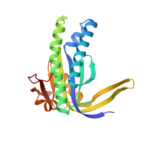
Crystal Structure and Regulation Mechanisms of the CyaB Adenylyl Cyclase from the Human Pathogen Pseudomonas aeruginosa.
Topal, H., Fulcher, N.B., Bitterman, J., Salazar, E., Buck, J., Levin, L.R., Cann, M.J., Wolfgang, M.C., Steegborn, C.(2012) J Mol Biology 416: 271-286
- PubMed: 22226839 Search on PubMedSearch on PubMed Central
- DOI: https://doi.org/10.1016/j.jmb.2011.12.045
- Primary Citation Related Structures:
3R5G - PubMed Abstract:
Pseudomonas aeruginosa is an opportunistic bacterial pathogen and a major cause of healthcare-associated infections. While the organism's intrinsic and acquired resistance to most antibiotics hinders treatment of P. aeruginosa infections, the regulatory networks controlling its virulence provide novel targets for drug development. CyaB, a key regulator of P. aeruginosa virulence, belongs to the Class III adenylyl cyclase (AC) family of enzymes that synthesize the second messenger cyclic adenosine 3',5'-monophosphate. These enzymes consist of a conserved catalytic domain fused to one or more regulatory domains. We describe here the biochemical and structural characterization of CyaB and its inhibition by small molecules. We show that CyaB belongs to the Class IIIb subfamily, and like other subfamily members, its activity is stimulated by inorganic carbon. CyaB is also regulated by its N-terminal MASE2 (membrane-associated sensor 2) domain, which acts as a membrane anchor. Using a genetic screen, we identified activating mutations in CyaB. By solving the crystal structure of the CyaB catalytic domain, we rationalized the effects of these mutations and propose that CyaB employs regulatory mechanisms similar to other Class III ACs. The CyaB structure further indicates subtle differences compared to other Class III ACs in both the active site and the inhibitor binding pocket. Consistent with these differences, we observed a unique inhibition profile, including identification of a CyaB selective compound. Overall, our results reveal mechanistic details of the physiological and pharmacological regulation of CyaB and provide the basis for its exploitation as a therapeutic drug target.
- Department of Physiological Chemistry, Ruhr-University Bochum, 44801 Bochum, Germany.
Organizational Affiliation: