3R4L

Human very long half life Plasminogen Activator Inhibitor type-1


Experimental Data Snapshot

  • Method: X-RAY DIFFRACTION
  • Resolution: 2.70 Å
  • R-Value Free: 0.282 
  • R-Value Work: 0.209 
  • R-Value Observed: 0.212 

wwPDB Validation   3D Report Full Report


Ligand Structure Quality Assessment 


This is version 2.1 of the entry. See complete history


Literature

Remarkable extension of PAI-1 half-life surprisingly brings no changes to its structure.

Jankun, J.Yang, J.Zheng, H.Han, F.Q.Al-Senaidy, A.Skrzypczak-Jankun, E.

(2012) Int J Mol Med 29: 61-64

  • DOI: https://doi.org/10.3892/ijmm.2011.798
  • Primary Citation of Related Structures:  
    3R4L

  • PubMed Abstract: 

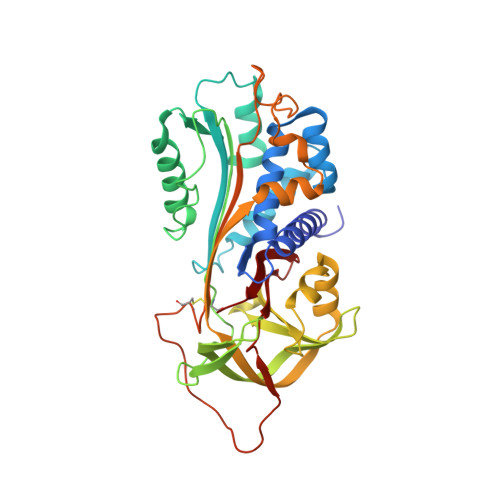
    Plasminogen activator inhibitor type 1 (PAI-1) is a serpin protein, a natural inhibitor of urokinase (uPA) and tissue plasminogen activators (tPA). By inhibiting uPA it can block growth of the cancer tumors by suppressing angiogenesis, while when acting on tPA in the blood it can avert conversion of plasminogen to plasmin preventing lysis of the clot. Furthermore, blocking PAI-1 activity can protect against thrombosis. Thus PAI-1 makes great impact on human homeostasis and is desirable for clinical application. Wild-type PAI-1 (wt-PAI-1) has a short span of activity with a t1/2 of ~2 h, being spontaneously converted into a latent form. An enormous effort has been made to create a more stable molecule with >600 PAI-1 variants constructed to study its structure-function relationship. In the present study, we evaluate the structure of the active recombinant VLHL-PAI-1 (very long half life, active >700 h) which is glycosylated similarly to wt-PAI-1 at N232 and N288, with the extended reactive center loop, intact engineered -S-S-bridge (Q174C, G323C) that precludes latency without affecting structure, and can be controlled by a reducing agent to terminate activity at will. We have already proven its usefulness to control cancer in human cancer cells, as well as preventing clot lysis in human whole blood and plasma and in a mouse model. Our results demonstrate the potential therapeutic applications (topical or systemic) of this protein in the treatment of cancer, for the trauma patients to ward off an excessive blood loss, or for people with the PAI-1 deficiency, especially during surgery.


  • Organizational Affiliation

    Urology Research Center, Department of Urology, The University of Toledo, Toledo, OH 43614, USA. jerzy.jankun@utoledo.edu


Macromolecules
Find similar proteins by:  (by identity cutoff)  |  3D Structure
Entity ID: 1
MoleculeChains Sequence LengthOrganismDetailsImage
Plasminogen activator inhibitor 1379Homo sapiensMutation(s): 2 
Gene Names: PAI1PLANH1SERPINE1
UniProt & NIH Common Fund Data Resources
Find proteins for P05121 (Homo sapiens)
Explore P05121 
Go to UniProtKB:  P05121
PHAROS:  P05121
GTEx:  ENSG00000106366 
Entity Groups  
Sequence Clusters30% Identity50% Identity70% Identity90% Identity95% Identity100% Identity
UniProt GroupP05121
Sequence Annotations
Expand
  • Reference Sequence
Oligosaccharides

Help

Entity ID: 2
MoleculeChains Length2D Diagram Glycosylation3D Interactions
beta-D-mannopyranose-(1-6)-beta-D-mannopyranose-(1-4)-2-acetamido-2-deoxy-beta-D-glucopyranose-(1-4)-[alpha-L-fucopyranose-(1-6)]2-acetamido-2-deoxy-beta-D-glucopyranose
B
5N-Glycosylation
Glycosylation Resources
GlyTouCan:  G40247UV
GlyCosmos:  G40247UV
GlyGen:  G40247UV
Small Molecules
Ligands 1 Unique
IDChains Name / Formula / InChI Key2D Diagram3D Interactions
NAG
Query on NAG

Download Ideal Coordinates CCD File 
C [auth A]2-acetamido-2-deoxy-beta-D-glucopyranose
C8 H15 N O6
OVRNDRQMDRJTHS-FMDGEEDCSA-N
Experimental Data & Validation

Experimental Data

  • Method: X-RAY DIFFRACTION
  • Resolution: 2.70 Å
  • R-Value Free: 0.282 
  • R-Value Work: 0.209 
  • R-Value Observed: 0.212 
  • Space Group: P 31 2 1
Unit Cell:
Length ( Å )Angle ( ˚ )
a = 72.007α = 90
b = 72.007β = 90
c = 194.481γ = 120
Software Package:
Software NamePurpose
ADSCdata collection
MOLREPphasing
REFMACrefinement
DENZOdata reduction
SCALEPACKdata scaling

Structure Validation

View Full Validation Report


Ligand Structure Quality Assessment 


Entry History 

Deposition Data

Revision History  (Full details and data files)

  • Version 1.0: 2011-11-02
    Type: Initial release
  • Version 1.1: 2017-11-08
    Changes: Refinement description
  • Version 2.0: 2020-07-29
    Type: Remediation
    Reason: Carbohydrate remediation
    Changes: Advisory, Atomic model, Data collection, Database references, Derived calculations, Non-polymer description, Structure summary
  • Version 2.1: 2023-09-13
    Changes: Data collection, Database references, Refinement description, Structure summary