3R48

Crystal structure of a hetero-hexamer coiled coil


Experimental Data Snapshot

  • Method: X-RAY DIFFRACTION
  • Resolution: 2.00 Å
  • R-Value Free: 0.277 
  • R-Value Work: 0.210 
  • R-Value Observed: 0.213 

wwPDB Validation   3D Report Full Report


This is version 1.4 of the entry. See complete history


Literature

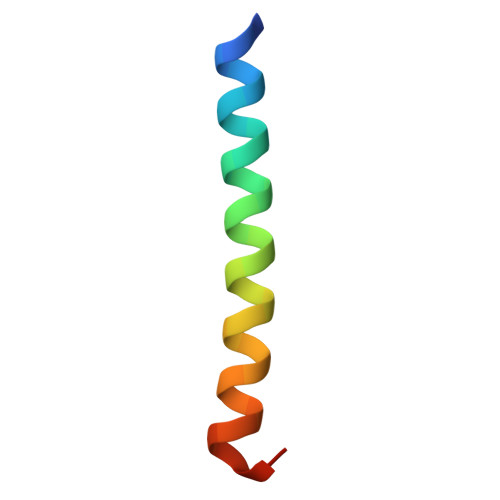
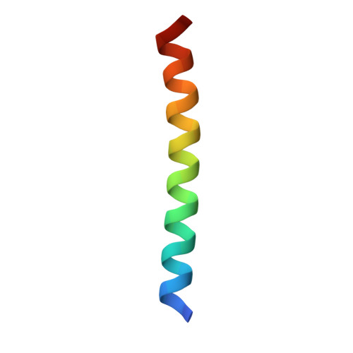
A de novo peptide hexamer with a mutable channel.

Zaccai, N.R.Chi, B.Thomson, A.R.Boyle, A.L.Bartlett, G.J.Bruning, M.Linden, N.Sessions, R.B.Booth, P.J.Brady, R.L.Woolfson, D.N.

(2011) Nat Chem Biol 7: 935-941

  • DOI: https://doi.org/10.1038/nchembio.692
  • Primary Citation of Related Structures:  
    3R3K, 3R46, 3R47, 3R48, 3R4A, 3R4H

  • PubMed Abstract: 

    The design of new proteins that expand the repertoire of natural protein structures represents a formidable challenge. Success in this area would increase understanding of protein structure and present new scaffolds that could be exploited in biotechnology and synthetic biology. Here we describe the design, characterization and X-ray crystal structure of a new coiled-coil protein. The de novo sequence forms a stand-alone, parallel, six-helix bundle with a channel running through it. Although lined exclusively by hydrophobic leucine and isoleucine side chains, the 6-Å channel is permeable to water. One layer of leucine residues within the channel is mutable, accepting polar aspartic acid and histidine side chains, which leads to subdivision and organization of solvent within the lumen. Moreover, these mutants can be combined to form a stable and unique (Asp-His)(3) heterohexamer. These new structures provide a basis for engineering de novo proteins with new functions.


  • Organizational Affiliation

    School of Biochemistry, University of Bristol, Bristol, UK.


Macromolecules
Find similar proteins by:  (by identity cutoff)  |  3D Structure
Entity ID: 1
MoleculeChains Sequence LengthOrganismDetailsImage
coiled coil helix W22-L24HA,
E [auth F],
F [auth G]
34synthetic constructMutation(s): 0 
Entity Groups  
Sequence Clusters30% Identity50% Identity70% Identity90% Identity95% Identity100% Identity
Sequence Annotations
Expand
  • Reference Sequence
Find similar proteins by:  (by identity cutoff)  |  3D Structure
Entity ID: 2
MoleculeChains Sequence LengthOrganismDetailsImage
coiled coil helix Y15-L24DB,
C,
D [auth E]
32synthetic constructMutation(s): 0 
Entity Groups  
Sequence Clusters30% Identity50% Identity70% Identity90% Identity95% Identity100% Identity
Sequence Annotations
Expand
  • Reference Sequence
Small Molecules
Ligands 1 Unique
IDChains Name / Formula / InChI Key2D Diagram3D Interactions
GOL
Query on GOL

Download Ideal Coordinates CCD File 
G [auth E]GLYCEROL
C3 H8 O3
PEDCQBHIVMGVHV-UHFFFAOYSA-N
Experimental Data & Validation

Experimental Data

  • Method: X-RAY DIFFRACTION
  • Resolution: 2.00 Å
  • R-Value Free: 0.277 
  • R-Value Work: 0.210 
  • R-Value Observed: 0.213 
  • Space Group: C 1 2 1
Unit Cell:
Length ( Å )Angle ( ˚ )
a = 148.872α = 90
b = 50.85β = 91.05
c = 31.681γ = 90
Software Package:
Software NamePurpose
PHENIXrefinement
SCALEPACKdata scaling
PHASERphasing
CNSrefinement
PDB_EXTRACTdata extraction
DENZOdata reduction

Structure Validation

View Full Validation Report


Entry History 

Deposition Data

Revision History  (Full details and data files)

  • Version 1.0: 2011-11-16
    Type: Initial release
  • Version 1.1: 2011-11-30
    Changes: Database references
  • Version 1.2: 2017-10-25
    Changes: Author supporting evidence
  • Version 1.3: 2019-07-17
    Changes: Data collection, Derived calculations, Refinement description, Source and taxonomy
  • Version 1.4: 2023-09-13
    Changes: Data collection, Database references, Derived calculations, Refinement description