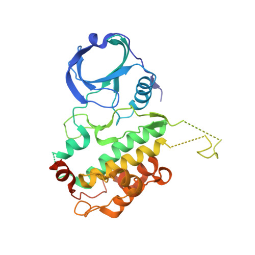
3R2B

MK2 kinase bound to Compound 5b


Experimental Data Snapshot

  • Method: X-RAY DIFFRACTION
  • Resolution: 2.90 Å
  • R-Value Free: 0.316 
  • R-Value Work: 0.277 
  • R-Value Observed: 0.279 

wwPDB Validation   3D Report Full Report


Ligand Structure Quality Assessment 


This is version 1.2 of the entry. See complete history


Literature

Structure-based lead identification of ATP-competitive MK2 inhibitors.

Barf, T.Kaptein, A.Wilde, S.Heijden, R.Someren, R.Demont, D.Schultz-Fademrecht, C.Versteegh, J.Zeeland, M.Seegers, N.Kazemier, B.Kar, B.Hoek, M.Roos, J.Klop, H.Smeets, R.Hofstra, C.Hornberg, J.Oubrie, A.

(2011) Bioorg Med Chem Lett 21: 3818-3822

  • DOI: https://doi.org/10.1016/j.bmcl.2011.04.018
  • Primary Citation of Related Structures:  
    3R1N, 3R2B, 3R2Y, 3R30

  • PubMed Abstract: 

    MK2 kinase is a promising drug discovery target for the treatment of inflammatory diseases. Here, we describe the discovery of novel MK2 inhibitors using X-ray crystallography and structure-based drug design. The lead has in vivo efficacy in a short-term preclinical model.


  • Organizational Affiliation

    Merck Research Laboratories, MSD, Oss, The Netherlands.


Macromolecules
Find similar proteins by:  (by identity cutoff)  |  3D Structure
Entity ID: 1
MoleculeChains Sequence LengthOrganismDetailsImage
MAP kinase-activated protein kinase 2
A, B, C, D, E
A, B, C, D, E, F, G, H, I, J, K, L
318Homo sapiensMutation(s): 0 
Gene Names: MAPKAPK2
EC: 2.7.11.1
UniProt & NIH Common Fund Data Resources
Find proteins for P49137 (Homo sapiens)
Explore P49137 
Go to UniProtKB:  P49137
PHAROS:  P49137
GTEx:  ENSG00000162889 
Entity Groups  
Sequence Clusters30% Identity50% Identity70% Identity90% Identity95% Identity100% Identity
UniProt GroupP49137
Sequence Annotations
Expand
  • Reference Sequence
Small Molecules
Ligands 1 Unique
IDChains Name / Formula / InChI Key2D Diagram3D Interactions
05B
Query on 05B

Download Ideal Coordinates CCD File 
M [auth A],
N [auth B]
2'-[2-(1,3-benzodioxol-5-yl)pyrimidin-4-yl]-5',6'-dihydrospiro[piperidine-4,7'-pyrrolo[3,2-c]pyridin]-4'(1'H)-one
C22 H21 N5 O3
QSDGMDKMSSYMKU-UHFFFAOYSA-N
Binding Affinity Annotations 
IDSourceBinding Affinity
05B PDBBind:  3R2B IC50: 4 (nM) from 1 assay(s)
BindingDB:  3R2B EC50: min: 4, max: 620 (nM) from 3 assay(s)
Experimental Data & Validation

Experimental Data

  • Method: X-RAY DIFFRACTION
  • Resolution: 2.90 Å
  • R-Value Free: 0.316 
  • R-Value Work: 0.277 
  • R-Value Observed: 0.279 
  • Space Group: P 21 21 21
Unit Cell:
Length ( Å )Angle ( ˚ )
a = 139.803α = 90
b = 180.249β = 90
c = 217.109γ = 90
Software Package:
Software NamePurpose
d*TREKdata scaling
REFMACrefinement
PDB_EXTRACTdata extraction
CrystalCleardata collection
d*TREKdata reduction
REFMACphasing

Structure Validation

View Full Validation Report


Ligand Structure Quality Assessment 


Entry History 

Deposition Data

Revision History  (Full details and data files)

  • Version 1.0: 2011-05-25
    Type: Initial release
  • Version 1.1: 2011-07-13
    Changes: Version format compliance
  • Version 1.2: 2024-02-21
    Changes: Data collection, Database references, Derived calculations, Refinement description