3R1A

Closed crystal structure of cytochrome P450 2B4 covalently bound to the mechanism-based inactivator tert-butylphenylacetylene


Experimental Data Snapshot

  • Method: X-RAY DIFFRACTION
  • Resolution: 3.50 Å
  • R-Value Free: 0.285 
  • R-Value Work: 0.206 
  • R-Value Observed: 0.210 

wwPDB Validation   3D Report Full Report


Ligand Structure Quality Assessment 


This is version 1.2 of the entry. See complete history


Literature

Structural Analysis of Mammalian Cytochrome P450 2B4 Covalently Bound to the Mechanism-Based Inactivator tert-Butylphenylacetylene: Insight into Partial Enzymatic Activity.

Gay, S.C.Zhang, H.Wilderman, P.R.Roberts, A.G.Liu, T.Li, S.Lin, H.L.Zhang, Q.Woods, V.L.Stout, C.D.Hollenberg, P.F.Halpert, J.R.

(2011) Biochemistry 50: 4903-4911

  • DOI: https://doi.org/10.1021/bi200482g
  • Primary Citation of Related Structures:  
    3R1A

  • PubMed Abstract: 

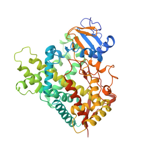
    A combined structural and computational analysis of rabbit cytochrome P450 2B4 covalently bound to the mechanism-based inactivator tert-butylphenylacetylene (tBPA) has yielded insight into how the enzyme retains partial activity. Since conjugation to tBPA modifies a highly conserved active site residue, the residual activity of tBPA-labeled 2B4 observed in previous studies was puzzling. Here we describe the first crystal structures of a modified mammalian P450, which show an oxygenated metabolite of tBPA conjugated to Thr 302 of helix I. These results are consistent with previous studies that identified Thr 302 as the site of conjugation. In each structure, the core of 2B4 remains unchanged, but the arrangement of plastic regions differs. This results in one structure that is compact and closed. In this conformation, tBPA points toward helix B', making a 31° angle with the heme plane. This conformation is in agreement with previously performed in silico experiments. However, dimerization of 2B4 in the other structure, which is caused by movement of the B/C loop and helices F through G, alters the position of tBPA. In this case, tBPA lies almost parallel to the heme plane due to the presence of helix F' of the opposite monomer entering the active site to stabilize the dimer. However, docking experiments using this open form show that tBPA is able to rotate upward to give testosterone and 7-ethoxy-4-trifluoromethylcoumarin access to the heme, which could explain the previously observed partial activity.


  • Organizational Affiliation

    Skaggs School of Pharmacy and Pharmaceutical Sciences, University of California, San Diego, La Jolla, California 92093, USA. scgay@ucsd.edu


Macromolecules
Find similar proteins by:  (by identity cutoff)  |  3D Structure
Entity ID: 1
MoleculeChains Sequence LengthOrganismDetailsImage
Cytochrome P450 2B4
A, B, C, D, E
A, B, C, D, E, F, G, H
476Oryctolagus cuniculusMutation(s): 9 
Gene Names: CYP2B4
EC: 1.14.14.1
UniProt
Find proteins for P00178 (Oryctolagus cuniculus)
Explore P00178 
Go to UniProtKB:  P00178
Entity Groups  
Sequence Clusters30% Identity50% Identity70% Identity90% Identity95% Identity100% Identity
UniProt GroupP00178
Sequence Annotations
Expand
  • Reference Sequence
Small Molecules
Ligands 2 Unique
IDChains Name / Formula / InChI Key2D Diagram3D Interactions
HEM
Query on HEM

Download Ideal Coordinates CCD File 
I [auth A]
K [auth B]
M [auth C]
O [auth D]
Q [auth E]
I [auth A],
K [auth B],
M [auth C],
O [auth D],
Q [auth E],
S [auth F],
U [auth G],
W [auth H]
PROTOPORPHYRIN IX CONTAINING FE
C34 H32 Fe N4 O4
KABFMIBPWCXCRK-RGGAHWMASA-L
TB2
Query on TB2

Download Ideal Coordinates CCD File 
J [auth A]
L [auth B]
N [auth C]
P [auth D]
R [auth E]
J [auth A],
L [auth B],
N [auth C],
P [auth D],
R [auth E],
T [auth F],
V [auth G],
X [auth H]
(4-tert-butylphenyl)acetaldehyde
C12 H16 O
VMLYBYNXKMHLIJ-UHFFFAOYSA-N
Experimental Data & Validation

Experimental Data

  • Method: X-RAY DIFFRACTION
  • Resolution: 3.50 Å
  • R-Value Free: 0.285 
  • R-Value Work: 0.206 
  • R-Value Observed: 0.210 
  • Space Group: P 21 21 21
Unit Cell:
Length ( Å )Angle ( ˚ )
a = 134.381α = 90
b = 144.549β = 90
c = 229.275γ = 90
Software Package:
Software NamePurpose
MOSFLMdata reduction
SCALAdata scaling
PHASERphasing
PHENIXrefinement
PDB_EXTRACTdata extraction
SSRLdata collection

Structure Validation

View Full Validation Report


Ligand Structure Quality Assessment 


Entry History 

Deposition Data

Revision History  (Full details and data files)

  • Version 1.0: 2011-05-11
    Type: Initial release
  • Version 1.1: 2011-07-13
    Changes: Version format compliance
  • Version 1.2: 2023-09-13
    Changes: Data collection, Database references, Derived calculations, Refinement description