


The Crystal Structure of human Histone-lysine N-methyltransferase SMYD3
Dombrovski, L., Dong, A., Wu, H., Min, J.To be published.
Experimental Data Snapshot
Starting Model: experimental
View more details
Entity ID: 1 | |||||
|---|---|---|---|---|---|
| Molecule | Chains | Sequence Length | Organism | Details | Image |
| SET and MYND domain-containing protein 3 | 429 | Homo sapiens | Mutation(s): 0 Gene Names: SMYD3, ZMYND1, ZNFN3A1 EC: 2.1.1.43 (PDB Primary Data), 2.1.1.354 (UniProt) | 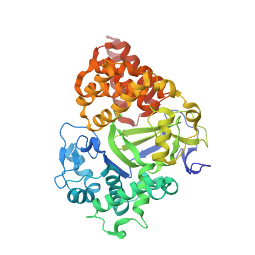 | |
UniProt & NIH Common Fund Data Resources | |||||
PHAROS: Q9H7B4 GTEx: ENSG00000185420 | |||||
Entity Groups | |||||
| Sequence Clusters | 30% Identity50% Identity70% Identity90% Identity95% Identity100% Identity | ||||
| UniProt Group | Q9H7B4 | ||||
Sequence AnnotationsExpand | |||||
Reference Sequence | |||||
| Ligands 3 Unique | |||||
|---|---|---|---|---|---|
| ID | Chains | Name / Formula / InChI Key | 2D Diagram | 3D Interactions | |
| SAM Download:Ideal Coordinates CCD File | E [auth A] | S-ADENOSYLMETHIONINE C15 H22 N6 O5 S MEFKEPWMEQBLKI-FCKMPRQPSA-N |  | ||
| GOL Download:Ideal Coordinates CCD File | F [auth A] G [auth A] H [auth A] I [auth A] J [auth A] | GLYCEROL C3 H8 O3 PEDCQBHIVMGVHV-UHFFFAOYSA-N |  | ||
| ZN Download:Ideal Coordinates CCD File | B [auth A], C [auth A], D [auth A] | ZINC ION Zn PTFCDOFLOPIGGS-UHFFFAOYSA-N |  | ||
| Length ( Å ) | Angle ( ˚ ) |
|---|---|
| a = 61.303 | α = 90 |
| b = 66.206 | β = 90 |
| c = 107.387 | γ = 90 |
| Software Name | Purpose |
|---|---|
| HKL-3000 | data collection |
| PHASER | phasing |
| REFMAC | refinement |
| Coot | model building |
| HKL-3000 | data reduction |
| HKL-3000 | data scaling |


