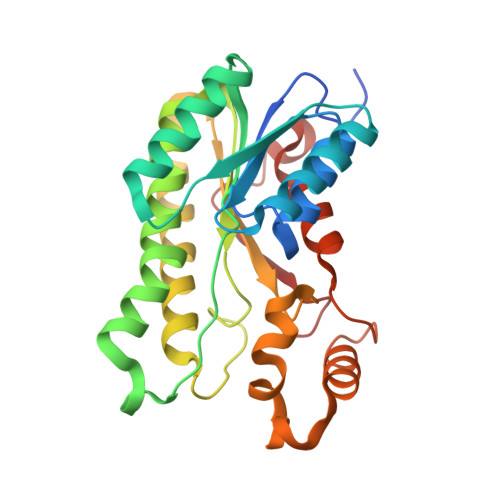
Structural basis for inhibition of 17 beta-hydroxysteroid dehydrogenases by phytoestrogens: The case of fungal 17 beta-HSDcl.
Cassetta, A., Stojan, J., Krastanova, I., Kristan, K., Brunskole Svegelj, M., Lamba, D., Rizner, T.L.(2017) J Steroid Biochem Mol Biol 171: 80-93
- PubMed: 28259640 Search on PubMed
- DOI: https://doi.org/10.1016/j.jsbmb.2017.02.020
- Primary Citation Related Structures:
3QWH, 4FJ0, 4FJ1, 4FJ2 - PubMed Abstract:
Phytoestrogens are plant-derived compounds that functionally and structurally mimic mammalian estrogens. Phytoestrogens have broad inhibitory activities toward several steroidogenic enzymes, such as the 17β-hydroxysteroid dehydrogenases (17β-HSDs), which modulate the biological potency of androgens and estrogens in mammals. However, to date, no crystallographic data are available to explain phytoestrogens binding to mammalian 17β-HSDs. NADP(H)-dependent 17β-HSD from the filamentous fungus Cochliobolus lunatus (17β-HSDcl) has been the subject of extensive biochemical, kinetic and quantitative structure-activity relationship studies that have shown that the flavonols are the most potent inhibitors. In the present study, we investigated the structure-activity relationships of the ternary complexes between the holo form of 17β-HSDcl and the flavonols kaempferol and 3,7-dihydroxyflavone, in comparison with the isoflavones genistein and biochanin A. Crystallographic data are accompanied by kinetic analysis of the inhibition mechanisms for six flavonols (3-hydroxyflavone, 3,7-dihydroxyflavone, kaempferol, quercetin, fisetin, myricetin), one flavanone (naringenin), one flavone (luteolin), and two isoflavones (genistein, biochanin A). The kinetics analysis shows that the degree of hydroxylation of ring B significantly influences the overall inhibitory efficacy of the flavonols. A distinct binding mode defines the interactions between 17β-HSDcl and the flavones and isoflavones. Moreover, the complex with biochanin A reveals an unusual binding mode that appears to account for its greater inhibition of 17β-HSDcl with respect to genistein. Overall, these data provide a blueprint for identification of the distinct molecular determinants that underpin 17β-HSD inhibition by phytoestrogens.
- Istituto di Cristallografia, UOS Trieste, Consiglio Nazionale delle Ricerche, S. S. 14-Km 163.5, I-34149, Trieste, Italy. Electronic address: alberto.cassetta@ts.ic.cnr.it.
Organizational Affiliation: