3QVA | pdb_00003qva

Structure of Klebsiella pneumoniae 5-hydroxyisourate hydrolase


Experimental Data Snapshot

  • Method: X-RAY DIFFRACTION
  • Resolution: 1.75 Å
  • R-Value Free: 
    0.223 (Depositor), 0.222 (DCC) 
  • R-Value Work: 
    0.199 (Depositor), 0.198 (DCC) 
  • R-Value Observed: 
    0.200 (Depositor) 

wwPDB Validation 3D Report Full Report

Validation slider image for 3QVA

This is version 1.2 of the entry. See complete history

Literature

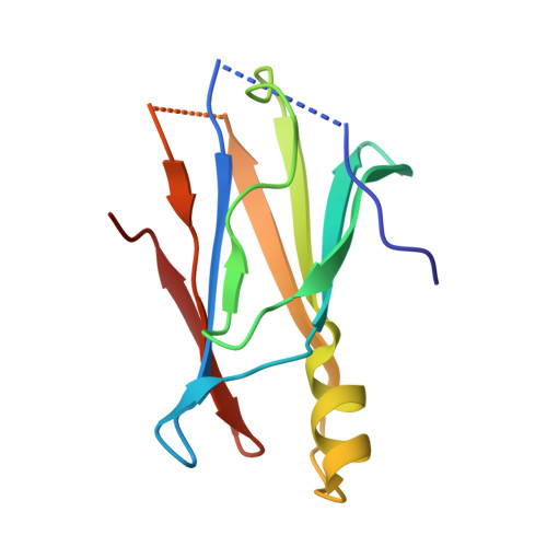
Structural and kinetic insights into the mechanism of 5-hydroxyisourate hydrolase from Klebsiella pneumoniae.

French, J.B.Ealick, S.E.

(2011) Acta Crystallogr D Biol Crystallogr 67: 671-677

  • DOI: https://doi.org/10.1107/S090744491101746X
  • Primary Citation Related Structures: 
    3QVA

  • PubMed Abstract: 

    The stereospecific oxidative degradation of uric acid to (S)-allantoin has recently been demonstrated to proceed via two unstable intermediates and requires three separate enzymatic reactions. The second step of this reaction, the conversion of 5-hydroxyisourate (HIU) to 2-oxo-4-hydroxy-4-carboxy-5-ureidoimidazoline, is catalyzed by HIU hydrolase (HIUH). The high-resolution crystal structure of HIUH from the opportunistic pathogen Klebsiella pneumoniae (KpHIUH) has been determined. KpHIUH is a homotetrameric protein that, based on sequence and structural similarity, belongs to the transthyretin-related protein family. In addition, the steady-state kinetic parameters for this enzyme and four active-site mutants have been measured. These data provide valuable insight into the functional roles of the active-site residues. Based upon the structural and kinetic data, a mechanism is proposed for the KpHIUH-catalyzed reaction.


  • Organizational Affiliation
    • Department of Chemistry and Chemical Biology, Cornell University, Ithaca, NY 14853-1301, USA.

Macromolecule Content 

  • Total Structure Weight: 49.96 kDa 
  • Atom Count: 3,454 
  • Modeled Residue Count: 422 
  • Deposited Residue Count: 464 
  • Unique protein chains: 1

Macromolecules

Find similar proteins by:|  3D Structure
Entity ID: 1
MoleculeChains  Sequence LengthOrganismDetailsImage
Transthyretin-like protein
A, B, C, D
116Klebsiella pneumoniae subsp. pneumoniae MGH 78578Mutation(s): 0 
Gene Names: HpxTKPN78578_16360KPN_01666
EC: 3.5.2.17
UniProt
Find proteins for A6T926 (Klebsiella pneumoniae subsp. pneumoniae (strain ATCC 700721 / MGH 78578))
Explore A6T926 
Go to UniProtKB:  A6T926
Entity Groups
Sequence Clusters30% Identity50% Identity70% Identity90% Identity95% Identity100% Identity
UniProt GroupA6T926
Sequence Annotations
Expand
Reference Sequence

Experimental Data & Validation

Experimental Data

  • Method: X-RAY DIFFRACTION
  • Resolution: 1.75 Å
  • R-Value Free:  0.223 (Depositor), 0.222 (DCC) 
  • R-Value Work:  0.199 (Depositor), 0.198 (DCC) 
  • R-Value Observed: 0.200 (Depositor) 
Space Group: P 21 21 21
Unit Cell:
Length ( Å )Angle ( ˚ )
a = 42.192α = 90
b = 101.302β = 90
c = 106.111γ = 90
Software Package:
Software NamePurpose
DENZOdata reduction
SCALEPACKdata scaling
MOLREPphasing
REFMACrefinement
PDB_EXTRACTdata extraction
HKL-2000data collection

Structure Validation

View Full Validation Report


Entry History 

Deposition Data

Revision History  (Full details and data files)

  • Version 1.0: 2011-08-24
    Type: Initial release
  • Version 1.1: 2017-11-08
    Changes: Refinement description
  • Version 1.2: 2024-02-21
    Changes: Data collection, Database references, Derived calculations