3QTP | pdb_00003qtp

Crystal Structure Analysis of Entamoeba histolytica Enolase


Experimental Data Snapshot

  • Method: X-RAY DIFFRACTION
  • Resolution: 1.90 Å
  • R-Value Free: 
    0.202 (Depositor), 0.200 (DCC) 
  • R-Value Work: 
    0.153 (Depositor), 0.154 (DCC) 
  • R-Value Observed: 
    0.155 (Depositor) 

wwPDB Validation 3D Report Full Report

Validation slider image for 3QTP

Ligand Structure Quality Assessment 


This is version 1.1 of the entry. See complete history

Literature

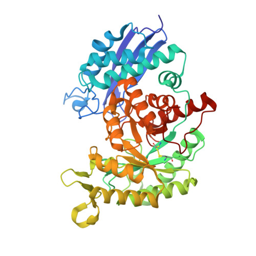
Structure analysis of Entamoeba histolytica enolase.

Schulz, E.C.Tietzel, M.Tovy, A.Ankri, S.Ficner, R.

(2011) Acta Crystallogr D Biol Crystallogr 67: 619-627

  • DOI: https://doi.org/10.1107/S0907444911016544
  • Primary Citation Related Structures: 
    3QTP

  • PubMed Abstract: 

    Entamoeba histolytica enolase (EhENO) reversibly interconverts 2-phosphoglyceric acid (2-PGA) and phosphoenolpyruvic acid (PEP). The crystal structure of the homodimeric EhENO is presented at a resolution of 1.9 Å. In the crystal structure EhENO presents as an asymmetric dimer with one active site in the open conformation and the other active site in the closed conformation. Interestingly, both active sites contain a copurified 2-PGA molecule. While the 2-PGA molecule in the closed active site closely resembles the conformation known from other enolase-2-PGA complexes, the conformation in the open active site is different. Here, 2-PGA is shifted approximately 1.6 Å away from metal ion I, most likely representing a precatalytic situation.


  • Organizational Affiliation
    • Abteilung für Molekulare Strukturbiologie, Institut für Mikrobiologie und Genetik, Georg-August-Universität Göttingen, Justus-von-Liebig Weg, Germany. eschulz1@gwdg.de

Macromolecule Content 

  • Total Structure Weight: 96.59 kDa 
  • Atom Count: 7,941 
  • Modeled Residue Count: 877 
  • Deposited Residue Count: 882 
  • Unique protein chains: 1

Macromolecules

Find similar proteins by:|  3D Structure
Entity ID: 1
MoleculeChains  Sequence LengthOrganismDetailsImage
Enolase 1
A, B
441Entamoeba histolyticaMutation(s): 0 
Gene Names: ENL-1
EC: 4.2.1.11
UniProt
Find proteins for P51555 (Entamoeba histolytica (strain ATCC 30459 / HM-1:IMSS / ABRM))
Explore P51555 
Go to UniProtKB:  P51555
Entity Groups
Sequence Clusters30% Identity50% Identity70% Identity90% Identity95% Identity100% Identity
UniProt GroupP51555
Sequence Annotations
Expand
Reference Sequence

Small Molecules

Ligands 3 Unique
IDChains Name / Formula / InChI Key2D Diagram3D Interactions
2PG

Query on 2PG


Download:Ideal Coordinates CCD File
C [auth A],
H [auth B]
2-PHOSPHOGLYCERIC ACID
C3 H7 O7 P
GXIURPTVHJPJLF-UWTATZPHSA-N
SO4

Query on SO4


Download:Ideal Coordinates CCD File
D [auth A]
E [auth A]
F [auth A]
I [auth B]
J [auth B]
D [auth A],
E [auth A],
F [auth A],
I [auth B],
J [auth B],
K [auth B]
SULFATE ION
O4 S
QAOWNCQODCNURD-UHFFFAOYSA-L
MG

Query on MG


Download:Ideal Coordinates CCD File
G [auth A],
L [auth B]
MAGNESIUM ION
Mg
JLVVSXFLKOJNIY-UHFFFAOYSA-N

Experimental Data & Validation

Experimental Data

  • Method: X-RAY DIFFRACTION
  • Resolution: 1.90 Å
  • R-Value Free:  0.202 (Depositor), 0.200 (DCC) 
  • R-Value Work:  0.153 (Depositor), 0.154 (DCC) 
  • R-Value Observed: 0.155 (Depositor) 
Space Group: P 21 21 21
Unit Cell:
Length ( Å )Angle ( ˚ )
a = 64.42α = 90
b = 92.62β = 90
c = 160.58γ = 90
Software Package:
Software NamePurpose
XSCALEdata scaling
PHASERphasing
REFMACrefinement
PDB_EXTRACTdata extraction
DNAdata collection
XDSdata reduction
PHENIXrefinement

Structure Validation

View Full Validation Report


Ligand Structure Quality Assessment 


Entry History 

Deposition Data

Revision History  (Full details and data files)

  • Version 1.0: 2011-07-13
    Type: Initial release
  • Version 1.1: 2024-10-30
    Changes: Data collection, Database references, Derived calculations, Structure summary