


Structure of Calcium Binding Protein-1 from Entamoeba histolytica in complex with Lead
Kumar, S., Gourinath, S.To be published.
Experimental Data Snapshot
Starting Model: experimental
View more details
wwPDB Validation 3D Report Full Report

Entity ID: 1 | |||||
|---|---|---|---|---|---|
| Molecule | Chains | Sequence Length | Organism | Details | Image |
| Calcium-binding protein | 134 | Entamoeba histolytica HM-1:IMSS | Mutation(s): 0 Gene Names: EhCaBP1 | 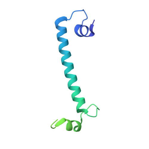 | |
UniProt | |||||
Entity Groups | |||||
| Sequence Clusters | 30% Identity50% Identity70% Identity90% Identity95% Identity100% Identity | ||||
| UniProt Group | P38505 | ||||
Sequence AnnotationsExpand | |||||
Reference Sequence | |||||
| Ligands 2 Unique | |||||
|---|---|---|---|---|---|
| ID | Chains | Name / Formula / InChI Key | 2D Diagram | 3D Interactions | |
| PB Download:Ideal Coordinates CCD File | C [auth A], D [auth A], F [auth B], G [auth B] | LEAD (II) ION Pb RVPVRDXYQKGNMQ-UHFFFAOYSA-N |  | ||
| ACT Download:Ideal Coordinates CCD File | E [auth A], H [auth B] | ACETATE ION C2 H3 O2 QTBSBXVTEAMEQO-UHFFFAOYSA-M |  | ||
| Length ( Å ) | Angle ( ˚ ) |
|---|---|
| a = 95.264 | α = 90 |
| b = 95.264 | β = 90 |
| c = 64.597 | γ = 120 |
| Software Name | Purpose |
|---|---|
| MAR345dtb | data collection |
| PHASER | phasing |
| CNS | refinement |
| AUTOMAR | data reduction |
| PHENIX | refinement |


