3QIP

Structure of HIV-1 reverse transcriptase in complex with an RNase H inhibitor and nevirapine


Experimental Data Snapshot

  • Method: X-RAY DIFFRACTION
  • Resolution: 2.09 Å
  • R-Value Free: 0.267 
  • R-Value Work: 0.216 
  • R-Value Observed: 0.219 

wwPDB Validation   3D Report Full Report


This is version 1.2 of the entry. See complete history


Literature

Structural and Binding Analysis of Pyrimidinol Carboxylic Acid and N-Hydroxy Quinazolinedione HIV-1 RNase H Inhibitors.

Lansdon, E.B.Liu, Q.Leavitt, S.A.Balakrishnan, M.Perry, J.K.Lancaster-Moyer, C.Kutty, N.Liu, X.Squires, N.H.Watkins, W.J.Kirschberg, T.A.

(2011) Antimicrob Agents Chemother 55: 2905-2915

  • DOI: https://doi.org/10.1128/AAC.01594-10
  • Primary Citation of Related Structures:  
    3QIN, 3QIO, 3QIP

  • PubMed Abstract: 

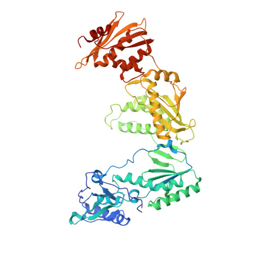
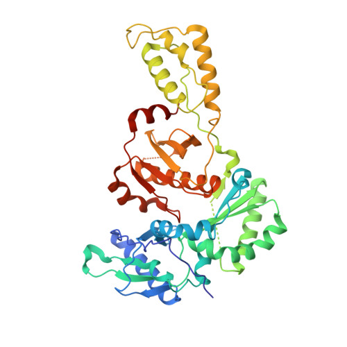
    HIV-1 RNase H breaks down the intermediate RNA-DNA hybrids during reverse transcription, requiring two divalent metal ions for activity. Pyrimidinol carboxylic acid and N-hydroxy quinazolinedione inhibitors were designed to coordinate the two metal ions in the active site of RNase H. High-resolution (1.4 Å to 2.1 Å) crystal structures were determined with the isolated RNase H domain and reverse transcriptase (RT), which permit accurate assessment of the metal and water environment at the active site. The geometry of the metal coordination suggests that the inhibitors mimic a substrate state prior to phosphodiester catalysis. Surface plasmon resonance studies confirm metal-dependent binding to RNase H and demonstrate that the inhibitors do not bind at the polymerase active site of RT. Additional evaluation of the RNase H site reveals an open protein surface with few additional interactions to optimize active-site inhibitors.


  • Organizational Affiliation

    Gilead Sciences, Inc., 333 Lakeside Dr., Foster City, CA 94404, USA. eric.lansdon@gilead.com


Macromolecules
Find similar proteins by:  (by identity cutoff)  |  3D Structure
Entity ID: 1
MoleculeChains Sequence LengthOrganismDetailsImage
Reverse HIV-1 reverse transcriptase p66560HIV-1 M:B_HXB2RMutation(s): 0 
Gene Names: gag-pol
EC: 3.4.23.16 (PDB Primary Data), 2.7.7.49 (PDB Primary Data), 2.7.7.7 (PDB Primary Data), 3.1.26.13 (PDB Primary Data), 3.1.13.2 (UniProt), 2.7.7 (UniProt), 3.1 (UniProt)
UniProt
Find proteins for P04585 (Human immunodeficiency virus type 1 group M subtype B (isolate HXB2))
Explore P04585 
Go to UniProtKB:  P04585
Entity Groups  
Sequence Clusters30% Identity50% Identity70% Identity90% Identity95% Identity100% Identity
UniProt GroupP04585
Sequence Annotations
Expand
  • Reference Sequence
Find similar proteins by:  (by identity cutoff)  |  3D Structure
Entity ID: 2
MoleculeChains Sequence LengthOrganismDetailsImage
p51440HIV-1 M:B_HXB2RMutation(s): 0 
Gene Names: gag-pol
EC: 3.4.23.16 (PDB Primary Data), 2.7.7.49 (PDB Primary Data), 2.7.7.7 (PDB Primary Data), 3.1.26.13 (PDB Primary Data), 3.1.13.2 (UniProt), 2.7.7 (UniProt), 3.1 (UniProt)
UniProt
Find proteins for P04585 (Human immunodeficiency virus type 1 group M subtype B (isolate HXB2))
Explore P04585 
Go to UniProtKB:  P04585
Entity Groups  
Sequence Clusters30% Identity50% Identity70% Identity90% Identity95% Identity100% Identity
UniProt GroupP04585
Sequence Annotations
Expand
  • Reference Sequence
Small Molecules
Ligands 5 Unique
IDChains Name / Formula / InChI Key2D Diagram3D Interactions
P4Y
Query on P4Y

Download Ideal Coordinates CCD File 
I [auth A]5,6-dihydroxy-2-[(2-phenyl-1H-indol-3-yl)methyl]pyrimidine-4-carboxylic acid
C20 H15 N3 O4
JONQCFJPPWOWOJ-UHFFFAOYSA-N
NVP
Query on NVP

Download Ideal Coordinates CCD File 
C [auth A]11-CYCLOPROPYL-5,11-DIHYDRO-4-METHYL-6H-DIPYRIDO[3,2-B:2',3'-E][1,4]DIAZEPIN-6-ONE
C15 H14 N4 O
NQDJXKOVJZTUJA-UHFFFAOYSA-N
SO4
Query on SO4

Download Ideal Coordinates CCD File 
F [auth A],
G [auth A]
SULFATE ION
O4 S
QAOWNCQODCNURD-UHFFFAOYSA-L
MN
Query on MN

Download Ideal Coordinates CCD File 
D [auth A],
E [auth A]
MANGANESE (II) ION
Mn
WAEMQWOKJMHJLA-UHFFFAOYSA-N
CL
Query on CL

Download Ideal Coordinates CCD File 
H [auth A],
J [auth B]
CHLORIDE ION
Cl
VEXZGXHMUGYJMC-UHFFFAOYSA-M
Binding Affinity Annotations 
IDSourceBinding Affinity
P4Y PDBBind:  3QIP Kd: 129 (nM) from 1 assay(s)
Experimental Data & Validation

Experimental Data

  • Method: X-RAY DIFFRACTION
  • Resolution: 2.09 Å
  • R-Value Free: 0.267 
  • R-Value Work: 0.216 
  • R-Value Observed: 0.219 
  • Space Group: C 2 2 21
Unit Cell:
Length ( Å )Angle ( ˚ )
a = 118.898α = 90
b = 154.876β = 90
c = 153.079γ = 90
Software Package:
Software NamePurpose
BOSdata collection
PHASERphasing
PHENIXrefinement
HKL-2000data reduction
HKL-2000data scaling

Structure Validation

View Full Validation Report


Entry History 

Deposition Data

Revision History  (Full details and data files)

  • Version 1.0: 2011-04-20
    Type: Initial release
  • Version 1.1: 2011-07-13
    Changes: Version format compliance
  • Version 1.2: 2023-09-13
    Changes: Data collection, Database references, Derived calculations, Refinement description