3QDQ

Complex between 4-hydroxybutyrate CoA-transferase from Clostridium aminobutyricum and CoA


Experimental Data Snapshot

  • Method: X-RAY DIFFRACTION
  • Resolution: 2.60 Å
  • R-Value Free: 0.274 
  • R-Value Work: 0.218 
  • R-Value Observed: 0.221 

wwPDB Validation   3D Report Full Report


Ligand Structure Quality Assessment 


This is version 1.3 of the entry. See complete history


Literature

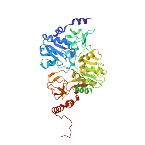
Crystal structure of the complex between 4-hydroxybutyrate CoA-transferase from Clostridium aminobutyricum and CoA.

Macieira, S.Zhang, J.Buckel, W.Messerschmidt, A.

(2012) Arch Microbiol 194: 157-166

  • DOI: https://doi.org/10.1007/s00203-011-0737-2
  • Primary Citation of Related Structures:  
    3QDQ

  • PubMed Abstract: 

    Clostridium aminobutyricum ferments 4-aminobutyrate (γ-aminobutyrate, GABA) to ammonia, acetate and butyrate via 4-hydroxybutyrate that is activated to the CoA-thioester catalyzed by 4-hydroxybutyrate CoA-transferase. Then, 4-hydroxybutyryl-CoA is dehydrated to crotonyl-CoA, which disproportionates to butyryl-CoA and acetyl-CoA. Cocrystallization of the CoA-transferase with the alternate substrate butyryl-CoA yielded crystals with non-covalently bound CoA and two water molecules at the active site. Most likely, butyryl-CoA reacted with the active site Glu238 to CoA and the mixed anhydride, which slowly hydrolyzed during crystallization. The structure of the CoA is similar but less stretched than that of the CoA-moiety of the covalent enzyme-CoA-thioester in 4-hydroxybutyrate CoA-transferase from Shewanella oneidensis. In contrast to the structures of the apo-enzyme and enzyme-CoA-thioester, the structure described here has a closed conformation, probably caused by a flip of the active site loop (residues 215-219). During turnover, the closed conformation may protect the anhydride intermediate from hydrolysis and CoA from dissociation from the enzyme. Hence, one catalytic cycle changes conformation of the enzyme four times: free enzyme-open conformation, CoA+ anhydride 1-closed, enzyme-CoA-thioester-open, CoA + anhydride-2-closed, free enzyme-open.


  • Organizational Affiliation

    Department of Proteomics and Signal Transduction, Max-Planck Institute of Biochemistry, Martinsried, Germany.


Macromolecules
Find similar proteins by:  (by identity cutoff)  |  3D Structure
Entity ID: 1
MoleculeChains Sequence LengthOrganismDetailsImage
4-Hydroxybutyrate CoA-transferase448Clostridium aminobutyricumMutation(s): 0 
Gene Names: abfT
EC: 2.8.3
UniProt
Find proteins for Q9RM86 (Clostridium aminobutyricum)
Explore Q9RM86 
Go to UniProtKB:  Q9RM86
Entity Groups  
Sequence Clusters30% Identity50% Identity70% Identity90% Identity95% Identity100% Identity
UniProt GroupQ9RM86
Sequence Annotations
Expand
  • Reference Sequence
Small Molecules
Ligands 1 Unique
IDChains Name / Formula / InChI Key2D Diagram3D Interactions
COA
Query on COA

Download Ideal Coordinates CCD File 
B [auth A]COENZYME A
C21 H36 N7 O16 P3 S
RGJOEKWQDUBAIZ-IBOSZNHHSA-N
Experimental Data & Validation

Experimental Data

  • Method: X-RAY DIFFRACTION
  • Resolution: 2.60 Å
  • R-Value Free: 0.274 
  • R-Value Work: 0.218 
  • R-Value Observed: 0.221 
  • Space Group: P 42 2 2
Unit Cell:
Length ( Å )Angle ( ˚ )
a = 118.215α = 90
b = 118.215β = 90
c = 87.392γ = 90
Software Package:
Software NamePurpose
MAR345data collection
MOLREPphasing
REFMACrefinement
HKL-2000data reduction
SCALEPACKdata scaling

Structure Validation

View Full Validation Report


Ligand Structure Quality Assessment 


Entry History 

Deposition Data

Revision History  (Full details and data files)

  • Version 1.0: 2011-08-24
    Type: Initial release
  • Version 1.1: 2012-02-29
    Changes: Database references
  • Version 1.2: 2017-11-08
    Changes: Refinement description
  • Version 1.3: 2023-09-13
    Changes: Data collection, Database references, Derived calculations, Refinement description