3QD7

Crystal structure of YdaL, a stand-alone small MutS-related protein from Escherichia coli


Experimental Data Snapshot

  • Method: X-RAY DIFFRACTION
  • Resolution: 2.30 Å
  • R-Value Free: 0.244 
  • R-Value Work: 0.224 
  • R-Value Observed: 0.225 

wwPDB Validation   3D Report Full Report


This is version 1.1 of the entry. See complete history


Literature

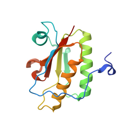
Crystal structure of YdaL, a stand-alone small MutS-related protein from Escherichia coli.

Gui, W.J.Qu, Q.H.Chen, Y.Y.Wang, M.Zhang, X.E.Bi, L.J.Jiang, T.

(2011) J Struct Biol 174: 282-289

  • DOI: https://doi.org/10.1016/j.jsb.2011.01.008
  • Primary Citation of Related Structures:  
    3QD7

  • PubMed Abstract: 
  • Sequence homologs of the small MutS-related (Smr) domain, the C-terminal endonuclease domain of MutS2, also exist as stand-alone proteins. In this study, we report the crystal structure of a proteolyzed fragment of YdaL (YdaL₃₉-₁₇₅), a stand-alone Smr protein from Escherichia coli ...

    Sequence homologs of the small MutS-related (Smr) domain, the C-terminal endonuclease domain of MutS2, also exist as stand-alone proteins. In this study, we report the crystal structure of a proteolyzed fragment of YdaL (YdaL₃₉-₁₇₅), a stand-alone Smr protein from Escherichia coli. In this structure, residues 86-170 assemble into a classical Smr core domain and are embraced by an N-terminal extension (residues 40-85) with an α/β/α fold. Sequence alignment indicates that the N-terminal extension is conserved among a number of stand-alone Smr proteins, suggesting structural diversity among Smr domains. We also discovered that the DNA binding affinity and endonuclease activity of the truncated YdaL₃₉-₁₇₅ protein were slightly lower than those of full-length YdaL₁-₁₈₇, suggesting that residues 1-38 may be involved in DNA binding.


    Organizational Affiliation

    National Laboratory of Biomacromolecules, Institute of Biophysics, Chinese Academy of Sciences, 15 Datun Road, Chaoyang District, Beijing 100101, China.


Macromolecules
Find similar proteins by:  (by identity cutoff)  |  3D Structure
Entity ID: 1
MoleculeChainsSequence LengthOrganismDetailsImage
Uncharacterized protein ydaLA [auth X]137Escherichia coli K-12Mutation(s): 0 
Gene Names: ydaLb1340JW1334smrA
EC: 3.1
UniProt
Find proteins for P76053 (Escherichia coli (strain K12))
Explore P76053 
Go to UniProtKB:  P76053
Entity Groups  
Sequence Clusters30% Identity50% Identity70% Identity90% Identity95% Identity100% Identity
UniProt GroupP76053
Protein Feature View
Expand
  • Reference Sequence
Experimental Data & Validation

Experimental Data

  • Method: X-RAY DIFFRACTION
  • Resolution: 2.30 Å
  • R-Value Free: 0.244 
  • R-Value Work: 0.224 
  • R-Value Observed: 0.225 
  • Space Group: P 21 21 2
Unit Cell:
Length ( Å )Angle ( ˚ )
a = 95.517α = 90
b = 34.833β = 90
c = 40.698γ = 90
Software Package:
Software NamePurpose
CrystalCleardata collection
PHENIXmodel building
REFMACrefinement
HKL-2000data reduction
SCALAdata scaling
PHENIXphasing

Structure Validation

View Full Validation Report


Entry History 

Deposition Data

Revision History  (Full details and data files)

  • Version 1.0: 2011-06-22
    Type: Initial release
  • Version 1.1: 2011-07-13
    Changes: Version format compliance