3PWY | pdb_00003pwy

Crystal structure of an extender (SPD28345)-modified human PDK1 complex 2


Experimental Data Snapshot

  • Method: X-RAY DIFFRACTION
  • Resolution: 3.50 Å
  • R-Value Free: 
    0.276 (Depositor), 0.271 (DCC) 
  • R-Value Work: 
    0.220 (Depositor), 0.210 (DCC) 
  • R-Value Observed: 
    0.226 (Depositor) 

Starting Model: experimental
View more details

wwPDB Validation 3D Report Full Report

Validation slider image for 3PWY

Ligand Structure Quality Assessment 


This is version 1.3 of the entry. See complete history

Literature

Discovery of a potent and highly selective PDK1 inhibitor via fragment-based drug discovery.

Erlanson, D.A.Arndt, J.W.Cancilla, M.T.Cao, K.Elling, R.A.English, N.Friedman, J.Hansen, S.K.Hession, C.Joseph, I.Kumaravel, G.Lee, W.C.Lind, K.E.McDowell, R.S.Miatkowski, K.Nguyen, C.Nguyen, T.B.Park, S.Pathan, N.Penny, D.M.Romanowski, M.J.Scott, D.Silvian, L.Simmons, R.L.Tangonan, B.T.Yang, W.Sun, L.

(2011) Bioorg Med Chem Lett 21: 3078-3083

  • DOI: https://doi.org/10.1016/j.bmcl.2011.03.032
  • Primary Citation Related Structures: 
    3PWY, 3QC4

  • PubMed Abstract: 

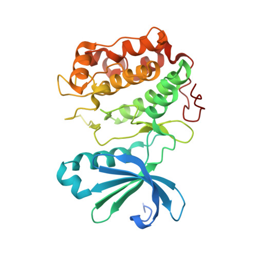
    We report the use of a fragment-based lead discovery method, Tethering with extenders, to discover a pyridinone fragment that binds in an adaptive site of the protein PDK1. With subsequent medicinal chemistry, this led to the discovery of a potent and highly selective inhibitor of PDK1, which binds in the 'DFG-out' conformation.


  • Organizational Affiliation
    • Sunesis Pharmaceuticals, Inc., 395 Oyster Point Blvd., South San Francisco, CA 94080, USA. derlanson@carmot.us

Macromolecule Content 

  • Total Structure Weight: 35.73 kDa 
  • Atom Count: 2,283 
  • Modeled Residue Count: 281 
  • Deposited Residue Count: 311 
  • Unique protein chains: 1

Macromolecules

Find similar proteins by:|  3D Structure
Entity ID: 1
MoleculeChains  Sequence LengthOrganismDetailsImage
3-phosphoinositide-dependent protein kinase 1311Homo sapiensMutation(s): 2 
Gene Names: PDK1PDPK1
EC: 2.7.11.1
UniProt & NIH Common Fund Data Resources
Find proteins for O15530 (Homo sapiens)
Explore O15530 
Go to UniProtKB:  O15530
PHAROS:  O15530
GTEx:  ENSG00000140992 
Entity Groups
Sequence Clusters30% Identity50% Identity70% Identity90% Identity95% Identity100% Identity
UniProt GroupO15530
Sequence Annotations
Expand
Reference Sequence

Small Molecules

Ligands 1 Unique
IDChains Name / Formula / InChI Key2D Diagram3D Interactions
SYP

Query on SYP


Download:Ideal Coordinates CCD File
B [auth A]N-[2-({6-[(2-sulfanylethyl)amino]pyrimidin-4-yl}amino)ethyl]propanamide
C11 H19 N5 O S
MXLYIGOSAJROJY-UHFFFAOYSA-N

Experimental Data & Validation

Experimental Data

  • Method: X-RAY DIFFRACTION
  • Resolution: 3.50 Å
  • R-Value Free:  0.276 (Depositor), 0.271 (DCC) 
  • R-Value Work:  0.220 (Depositor), 0.210 (DCC) 
  • R-Value Observed: 0.226 (Depositor) 
Space Group: P 32 2 1
Unit Cell:
Length ( Å )Angle ( ˚ )
a = 122.376α = 90
b = 122.376β = 90
c = 47.632γ = 120
Software Package:
Software NamePurpose
CrystalCleardata collection
MOLREPphasing
REFMACrefinement
MOSFLMdata reduction
SCALAdata scaling

Structure Validation

View Full Validation Report


Ligand Structure Quality Assessment 


Entry History 

Deposition Data

Revision History  (Full details and data files)

  • Version 1.0: 2011-04-20
    Type: Initial release
  • Version 1.1: 2011-07-13
    Changes: Version format compliance
  • Version 1.2: 2023-09-13
    Changes: Data collection, Database references, Derived calculations, Refinement description
  • Version 1.3: 2024-10-09
    Changes: Structure summary