3PPE | pdb_00003ppe

Crystal structure of chicken VE-cadherin EC1-2


Experimental Data Snapshot

  • Method: X-RAY DIFFRACTION
  • Resolution: 2.10 Å
  • R-Value Free: 
    0.242 (Depositor), 0.230 (DCC) 
  • R-Value Work: 
    0.184 (Depositor) 
  • R-Value Observed: 
    0.187 (Depositor) 

Starting Model: experimental
View more details

wwPDB Validation 3D Report Full Report

Validation slider image for 3PPE

This is version 1.2 of the entry. See complete history

Literature

Structure and binding mechanism of vascular endothelial cadherin: a divergent classical cadherin.

Brasch, J.Harrison, O.J.Ahlsen, G.Carnally, S.M.Henderson, R.M.Honig, B.Shapiro, L.

(2011) J Mol Biology 408: 57-73

  • DOI: https://doi.org/10.1016/j.jmb.2011.01.031
  • Primary Citation Related Structures: 
    3PPE

  • PubMed Abstract: 

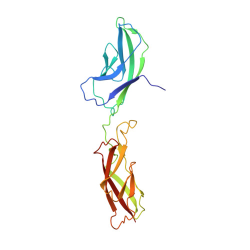
    Vascular endothelial cadherin (VE-cadherin), a divergent member of the type II classical cadherin family of cell adhesion proteins, mediates homophilic adhesion in the vascular endothelium. Previous investigations with a bacterially produced protein suggested that VE-cadherin forms cell surface trimers that bind between apposed cells to form hexamers. Here we report studies of mammalian-produced VE-cadherin ectodomains suggesting that, like other classical cadherins, VE-cadherin forms adhesive trans dimers between monomers located on opposing cell surfaces. Trimerization of the bacterially produced protein appears to be an artifact that arises from a lack of glycosylation. We also present the 2.1-Å-resolution crystal structure of the VE-cadherin EC1-2 adhesive region, which reveals homodimerization via the strand-swap mechanism common to classical cadherins. In common with type II cadherins, strand-swap binding involves two tryptophan anchor residues, but the adhesive interface resembles type I cadherins in that VE-cadherin does not form a large nonswapped hydrophobic surface. Thus, VE-cadherin is an outlier among classical cadherins, with characteristics of both type I and type II subfamilies.


  • Organizational Affiliation
    • Department of Biochemistry and Molecular Biophysics, Columbia University, 701 West 168th Street, New York, NY 10032, USA.

Macromolecule Content 

  • Total Structure Weight: 45.63 kDa 
  • Atom Count: 3,842 
  • Modeled Residue Count: 406 
  • Deposited Residue Count: 406 
  • Unique protein chains: 1

Macromolecules

Find similar proteins by:|  3D Structure
Entity ID: 1
MoleculeChains  Sequence LengthOrganismDetailsImage
Vascular endothelial cadherin
A, B
203Gallus gallusMutation(s): 0 
UniProt
Find proteins for Q8AYD0 (Gallus gallus)
Explore Q8AYD0 
Go to UniProtKB:  Q8AYD0
Entity Groups
Sequence Clusters30% Identity50% Identity70% Identity90% Identity95% Identity100% Identity
UniProt GroupQ8AYD0
Sequence Annotations
Expand
Reference Sequence

Experimental Data & Validation

Experimental Data

  • Method: X-RAY DIFFRACTION
  • Resolution: 2.10 Å
  • R-Value Free:  0.242 (Depositor), 0.230 (DCC) 
  • R-Value Work:  0.184 (Depositor) 
  • R-Value Observed: 0.187 (Depositor) 
Space Group: P 43 21 2
Unit Cell:
Length ( Å )Angle ( ˚ )
a = 99.973α = 90
b = 99.973β = 90
c = 105.987γ = 90
Software Package:
Software NamePurpose
MAR345dtbdata collection
PHASERphasing
REFMACrefinement
DENZOdata reduction
SCALEPACKdata scaling

Structure Validation

View Full Validation Report


Entry History 

Deposition Data

Revision History  (Full details and data files)

  • Version 1.0: 2011-02-02
    Type: Initial release
  • Version 1.1: 2011-07-13
    Changes: Version format compliance
  • Version 1.2: 2023-09-06
    Changes: Data collection, Database references, Derived calculations, Refinement description