Proline 96 of the copper ligand loop of amicyanin regulates electron transfer from methylamine dehydrogenase by positioning other residues at the protein-protein interface.
Choi, M., Sukumar, N., Mathews, F.S., Liu, A., Davidson, V.L.(2011) Biochemistry 50: 1265-1273
- PubMed: 21268585 Search on PubMedSearch on PubMed Central
- DOI: https://doi.org/10.1021/bi101794y
- Primary Citation Related Structures:
3PLY - PubMed Abstract:
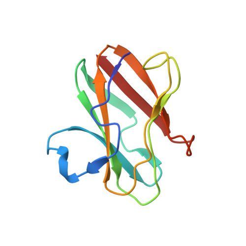
Amicyanin is a type 1 copper protein that serves as an electron acceptor for methylamine dehydrogenase (MADH). The site of interaction with MADH is a "hydrophobic patch" of amino acid residues including those that comprise a "ligand loop" that provides three of the four copper ligands. Three prolines are present in this region. Pro94 of the ligand loop was previously shown to strongly influence the redox potential of amicyanin but not affinity for MADH or mechanism of electron transfer (ET). In this study Pro96 of the ligand loop was mutated. P96A and P96G mutations did not affect the spectroscopic or redox properties of amicyanin but increased the K(d) for complex formation with MADH and altered the kinetic mechanism for the interprotein ET reaction. Values of reorganization energy (λ) and electronic coupling (H(AB)) for the ET reaction with MADH were both increased by the mutation, indicating that the true ET reaction observed with native amicyanin was now gated by or coupled to a reconfiguration of the proteins within the complex. The crystal structure of P96G amicyanin was very similar to that of native amicyanin, but notably, in addition to the change in Pro96, the side chains of residues Phe97 and Arg99 were oriented differently. These two residues were previously shown to make contacts with MADH that were important for stabilizing the amicyanin-MADH complex. The values of K(d), λ, and H(AB) for the reactions of the Pro96 mutants with MADH are remarkably similar to those obtained previously for P52G amicyanin. Mutation of this proline, also in the hydrophobic patch, caused reorientation of the side chain of Met51, another reside that interacted with MADH and caused a change in the kinetic mechanism of ET from MADH. These results show that proline residues near the copper site play key roles in positioning other amino acid residues at the amicyanin-MADH interface not only for specific binding to the redox protein partner but also to optimize the orientation of proteins for interprotein ET.
- Department of Biochemistry, University of Mississippi Medical Center, Jackson, Mississippi 39216, United States.
Organizational Affiliation: