3PL1

Determination of the crystal structure of the pyrazinamidase from M.tuberculosis : a structure-function analysis for prediction resistance to pyrazinamide.


Experimental Data Snapshot

  • Method: X-RAY DIFFRACTION
  • Resolution: 2.20 Å
  • R-Value Free: 0.240 
  • R-Value Work: 0.195 
  • R-Value Observed: 0.197 

wwPDB Validation   3D Report Full Report


This is version 1.3 of the entry. See complete history


Literature

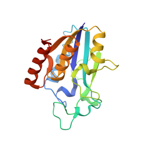
Crystal Structure of the Pyrazinamidase of Mycobacterium tuberculosis: Insights into Natural and Acquired Resistance to Pyrazinamide.

Petrella, S.Gelus-Ziental, N.Maudry, A.Laurans, C.Boudjelloul, R.Sougakoff, W.

(2011) PLoS One 6: e15785-e15785

  • DOI: https://doi.org/10.1371/journal.pone.0015785
  • Primary Citation of Related Structures:  
    3PL1

  • PubMed Abstract: 

    Pyrazinamidase (PncA) activates the first-line antituberculous drug pyrazinamide into pyrazinoic acid. The crystal structure of the Mycobacterium tuberculosis PncA protein has been determined, showing significant differences in the substrate binding cavity when compared to the pyrazinamidases from Pyrococcus horikoshii and Acinetobacter baumanii. In M. tuberculosis, this region was found to hold a Fe(2+) ion coordinated by one aspartate and three histidines, one of them corresponding to His57 which is replaced by Asp in Mycobacterium bovis, a species naturally resistant to pyrazinamide. The binding cavity also contains a Cys138-Asp8-Lys96 motif evocating a cysteine-based catalytic mechanism. Mutants have been constructed and investigated by kinetic and thermal shift assays, highlighting the importance of protein folding and thermal stability in the pyrazinamidase activity.


  • Organizational Affiliation

    Faculté de Médecine Pitié-Salpêtrière, UPMC Université Paris 6, ER 5, EA1541, Bactériologie-Hygiène, Paris, France.


Macromolecules
Find similar proteins by:  (by identity cutoff)  |  3D Structure
Entity ID: 1
MoleculeChains Sequence LengthOrganismDetailsImage
PYRAZINAMIDASE/NICOTINAMIDASE PNCA (PZase)186Mycobacterium tuberculosis H37RvMutation(s): 0 
Gene Names: MT2103pncARv2043c
EC: 3.5.1.19
UniProt
Find proteins for I6XD65 (Mycobacterium tuberculosis (strain ATCC 25618 / H37Rv))
Explore I6XD65 
Go to UniProtKB:  I6XD65
Entity Groups  
Sequence Clusters30% Identity50% Identity70% Identity90% Identity95% Identity100% Identity
UniProt GroupI6XD65
Sequence Annotations
Expand
  • Reference Sequence
Small Molecules
Ligands 1 Unique
IDChains Name / Formula / InChI Key2D Diagram3D Interactions
FE2
Query on FE2

Download Ideal Coordinates CCD File 
B [auth A]FE (II) ION
Fe
CWYNVVGOOAEACU-UHFFFAOYSA-N
Experimental Data & Validation

Experimental Data

  • Method: X-RAY DIFFRACTION
  • Resolution: 2.20 Å
  • R-Value Free: 0.240 
  • R-Value Work: 0.195 
  • R-Value Observed: 0.197 
  • Space Group: P 61 2 2
Unit Cell:
Length ( Å )Angle ( ˚ )
a = 84.1α = 90
b = 84.1β = 90
c = 100γ = 120
Software Package:
Software NamePurpose
PHASERphasing
REFMACrefinement
PDB_EXTRACTdata extraction
XDSdata reduction
XSCALEdata scaling

Structure Validation

View Full Validation Report


Entry History 

Deposition Data

Revision History  (Full details and data files)

  • Version 1.0: 2011-01-12
    Type: Initial release
  • Version 1.1: 2011-07-13
    Changes: Version format compliance
  • Version 1.2: 2014-11-12
    Changes: Structure summary
  • Version 1.3: 2023-11-01
    Changes: Data collection, Database references, Derived calculations, Refinement description