3PJS | pdb_00003pjs

Mechanism of Activation Gating in the Full-Length KcsA K+ Channel


Experimental Data Snapshot

  • Method: X-RAY DIFFRACTION
  • Resolution: 3.80 Å
  • R-Value Free: 
    0.332 (Depositor), 0.337 (DCC) 
  • R-Value Work: 
    0.281 (Depositor), 0.289 (DCC) 

wwPDB Validation 3D Report Full Report

Validation slider image for 3PJS

This is version 1.5 of the entry. See complete history

Literature

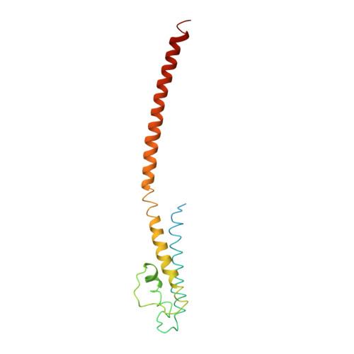
Mechanism of activation gating in the full-length KcsA K+ channel.

Uysal, S.Cuello, L.G.Cortes, D.M.Koide, S.Kossiakoff, A.A.Perozo, E.

(2011) Proc Natl Acad Sci U S A 108: 11896-11899

  • DOI: https://doi.org/10.1073/pnas.1105112108
  • Primary Citation Related Structures: 
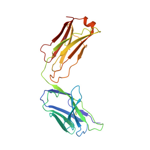
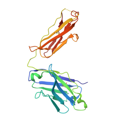
    3PJS

  • PubMed Abstract: 

    Using a constitutively active channel mutant, we solved the structure of full-length KcsA in the open conformation at 3.9 Å. The structure reveals that the activation gate expands about 20 Å, exerting a strain on the bulge helices in the C-terminal domain and generating side windows large enough to accommodate hydrated K(+) ions. Functional and spectroscopic analysis of the gating transition provides direct insight into the allosteric coupling between the activation gate and the selectivity filter. We show that the movement of the inner gate helix is transmitted to the C-terminus as a straightforward expansion, leading to an upward movement and the insertion of the top third of the bulge helix into the membrane. We suggest that by limiting the extent to which the inner gate can open, the cytoplasmic domain also modulates the level of inactivation occurring at the selectivity filter.


  • Organizational Affiliation
    • Department of Biochemistry and Molecular Biology, Institute for Biophysical Dynamics, University of Chicago, Chicago, IL 60637, USA.

Macromolecule Content 

  • Total Structure Weight: 168.97 kDa 
  • Atom Count: 10,982 
  • Modeled Residue Count: 1,424 
  • Deposited Residue Count: 1,542 
  • Unique protein chains: 3

Macromolecules

Find similar proteins by:|  3D Structure
Entity ID: 1
MoleculeChains  Sequence LengthOrganismDetailsImage
FAB light chain
A, C
215Mus musculusMutation(s): 0 
Find similar proteins by:|  3D Structure
Entity ID: 2
MoleculeChains  Sequence LengthOrganismDetailsImage
FAB heavy chain
B, D
224Mus musculusMutation(s): 0 
Find similar proteins by:|  3D Structure
Entity ID: 3
MoleculeChains  Sequence LengthOrganismDetailsImage
Voltage-gated potassium channelE [auth K],
F [auth L],
G [auth M],
H [auth N]
166Streptomyces lividansMutation(s): 7 
Gene Names: kcsAskc1
Membrane Entity: Yes 
UniProt
Find proteins for P0A334 (Streptomyces lividans)
Explore P0A334 
Go to UniProtKB:  P0A334
Entity Groups
Sequence Clusters30% Identity50% Identity70% Identity90% Identity95% Identity100% Identity
UniProt GroupP0A334
Sequence Annotations
Expand
Reference Sequence

Experimental Data & Validation

Experimental Data

  • Method: X-RAY DIFFRACTION
  • Resolution: 3.80 Å
  • R-Value Free:  0.332 (Depositor), 0.337 (DCC) 
  • R-Value Work:  0.281 (Depositor), 0.289 (DCC) 
Space Group: I 2 2 2
Unit Cell:
Length ( Å )Angle ( ˚ )
a = 118.266α = 90
b = 176.716β = 90
c = 340.466γ = 90
Software Package:
Software NamePurpose
MAR345dtbdata collection
PHASERphasing
CNSrefinement
HKL-2000data reduction
HKL-2000data scaling

Structure Validation

View Full Validation Report


Entry History 

Deposition Data

Revision History  (Full details and data files)

  • Version 1.0: 2011-07-06
    Type: Initial release
  • Version 1.1: 2011-07-13
    Changes: Version format compliance
  • Version 1.2: 2011-07-20
    Changes: Database references
  • Version 1.3: 2011-08-03
    Changes: Database references
  • Version 1.4: 2024-11-20
    Changes: Data collection, Database references, Structure summary
  • Version 1.5: 2026-01-28
    Changes: Derived calculations