Conformational change upon product binding to Klebsiella pneumoniae UDP-glucose dehydrogenase: a possible inhibition mechanism for the key enzyme in polymyxin resistance.
Chen, Y.Y., Ko, T.P., Lin, C.H., Chen, W.H., Wang, A.H.(2011) J Struct Biol 175: 300-310
- PubMed: 21536136 Search on PubMed
- DOI: https://doi.org/10.1016/j.jsb.2011.04.010
- Primary Citation Related Structures:
3PHL, 3PID, 3PJG, 3PLN, 3PLR - PubMed Abstract:
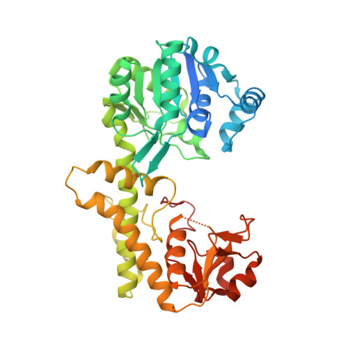
Cationic modification of lipid A with 4-amino-4-deoxy-L-arabinopyranose (L-Ara4N) allows the pathogen Klebsiella pneumoniae to resist the antibiotic polymyxin and other cationic antimicrobial peptides. UDP-glucose dehydrogenase (Ugd) catalyzes the NAD⁺-dependent twofold oxidation of UDP-glucose (UPG) to produce UDP-glucuronic acid (UGA), a requisite precursor in the biosynthesis of L-Ara4N and bacterial exopolysaccharides. Here we report five crystal structures of K. pneumoniae Ugd (KpUgd) in its apo form, in complex with UPG, UPG/NADH, two UGA molecules, and finally with a C-terminal His₆-tag. The UGA-complex structure differs from the others by a 14° rotation of the N-terminal domain toward the C-terminal domain, and represents a closed enzyme conformation. It also reveals that the second UGA molecule binds to a pre-existing positively charged surface patch away from the active site. The enzyme is thus inactivated by moving the catalytically important residues C253, K256 and D257 from their original positions. Kinetic data also suggest that KpUgd has multiple binding sites for UPG, and that UGA is a competitive inhibitor. The conformational changes triggered by UGA binding to the allosteric site can be exploited in designing potent inhibitors.
- Institute of Biological Chemistry, Academia Sinica, Taipei 115, Taiwan; Institute of Biochemical Sciences, National Taiwan University, Taipei 106, Taiwan.
Organizational Affiliation: