3PB1 | pdb_00003pb1

Crystal Structure of a Michaelis Complex between Plasminogen Activator Inhibitor-1 and Urokinase-type Plasminogen Activator


Experimental Data Snapshot

  • Method: X-RAY DIFFRACTION
  • Resolution: 2.30 Å
  • R-Value Free: 
    0.272 (Depositor), 0.269 (DCC) 
  • R-Value Work: 
    0.220 (Depositor), 0.217 (DCC) 
  • R-Value Observed: 
    0.223 (Depositor) 

Starting Model: experimental
View more details

wwPDB Validation 3D Report Full Report

Validation slider image for 3PB1

This is version 1.3 of the entry. See complete history

Literature

Structural basis for recognition of urokinase-type plasminogen activator by plasminogen activator inhibitor-1.

Lin, Z.Jiang, L.Yuan, C.Jensen, J.K.Zhang, X.Luo, Z.Furie, B.C.Furie, B.Andreasen, P.A.Huang, M.

(2011) J Biological Chem 286: 7027-7032

  • DOI: https://doi.org/10.1074/jbc.M110.204537
  • Primary Citation Related Structures: 
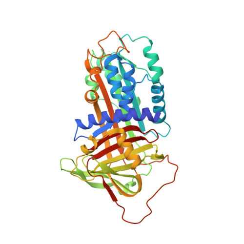
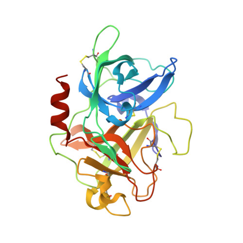
    3PB1

  • PubMed Abstract: 

    Plasminogen activator inhibitor-1 (PAI-1), together with its physiological target urokinase-type plasminogen activator (uPA), plays a pivotal role in fibrinolysis, cell migration, and tissue remodeling and is currently recognized as being among the most extensively validated biological prognostic factors in several cancer types. PAI-1 specifically and rapidly inhibits uPA and tissue-type PA (tPA). Despite extensive structural/functional studies on these two reactions, the underlying structural mechanism has remained unknown due to the technical difficulties of obtaining the relevant structures. Here, we report a strategy to generate a PAI-1·uPA(S195A) Michaelis complex and present its crystal structure at 2.3-Å resolution. In this structure, the PAI-1 reactive center loop serves as a bait to attract uPA onto the top of the PAI-1 molecule. The P4-P3' residues of the reactive center loop interact extensively with the uPA catalytic site, accounting for about two-thirds of the total contact area. Besides the active site, almost all uPA exosite loops, including the 37-, 60-, 97-, 147-, and 217-loops, are involved in the interaction with PAI-1. The uPA 37-loop makes an extensive interaction with PAI-1 β-sheet B, and the 147-loop directly contacts PAI-1 β-sheet C. Both loops are important for initial Michaelis complex formation. This study lays down a foundation for understanding the specificity of PAI-1 for uPA and tPA and provides a structural basis for further functional studies.


  • Organizational Affiliation
    • State Key Laboratory of Structural Chemistry, Fujian Institute of Research on the Structure of Matter, Chinese Academy of Sciences, Fuzhou 350002, China.

Macromolecule Content 

  • Total Structure Weight: 71.41 kDa 
  • Atom Count: 5,250 
  • Modeled Residue Count: 622 
  • Deposited Residue Count: 632 
  • Unique protein chains: 2

Macromolecules

Find similar proteins by:|  3D Structure
Entity ID: 1
MoleculeChains  Sequence LengthOrganismDetailsImage
Plasminogen activator inhibitor 1A [auth I]379Homo sapiensMutation(s): 4 
Gene Names: SERPINE1PAI1PLANH1
UniProt & NIH Common Fund Data Resources
Find proteins for P05121 (Homo sapiens)
Explore P05121 
Go to UniProtKB:  P05121
PHAROS:  P05121
GTEx:  ENSG00000106366 
Entity Groups
Sequence Clusters30% Identity50% Identity70% Identity90% Identity95% Identity100% Identity
UniProt GroupP05121
Sequence Annotations
Expand
Reference Sequence
Find similar proteins by:|  3D Structure
Entity ID: 2
MoleculeChains  Sequence LengthOrganismDetailsImage
Plasminogen activator, urokinaseB [auth E]253Homo sapiensMutation(s): 1 
Gene Names: PLAURP11-417O11.1-002
EC: 3.4.21.73
UniProt & NIH Common Fund Data Resources
Find proteins for P00749 (Homo sapiens)
Explore P00749 
Go to UniProtKB:  P00749
PHAROS:  P00749
GTEx:  ENSG00000122861 
Entity Groups
Sequence Clusters30% Identity50% Identity70% Identity90% Identity95% Identity100% Identity
UniProt GroupP00749
Sequence Annotations
Expand
Reference Sequence

Experimental Data & Validation

Experimental Data

  • Method: X-RAY DIFFRACTION
  • Resolution: 2.30 Å
  • R-Value Free:  0.272 (Depositor), 0.269 (DCC) 
  • R-Value Work:  0.220 (Depositor), 0.217 (DCC) 
  • R-Value Observed: 0.223 (Depositor) 
Space Group: P 32 2 1
Unit Cell:
Length ( Å )Angle ( ˚ )
a = 97.711α = 90
b = 97.711β = 90
c = 171.916γ = 120
Software Package:
Software NamePurpose
HKL-2000data collection
CCP4model building
REFMACrefinement
HKL-2000data reduction
HKL-2000data scaling
CCP4phasing

Structure Validation

View Full Validation Report


Entry History 

Deposition Data

Revision History  (Full details and data files)

  • Version 1.0: 2010-12-29
    Type: Initial release
  • Version 1.1: 2011-07-13
    Changes: Version format compliance
  • Version 1.2: 2023-11-01
    Changes: Data collection, Database references, Derived calculations, Refinement description
  • Version 1.3: 2024-11-06
    Changes: Structure summary