3P4P | pdb_00003p4p

Crystal structure of Menaquinol:fumarate oxidoreductase in complex with fumarate


Experimental Data Snapshot

  • Method: X-RAY DIFFRACTION
  • Resolution: 2.80 Å
  • R-Value Free: 
    0.272 (Depositor), 0.278 (DCC) 
  • R-Value Work: 
    0.249 (Depositor), 0.258 (DCC) 
  • R-Value Observed: 
    0.249 (Depositor) 

wwPDB Validation 3D Report Full Report

Validation slider image for 3P4P

Ligand Structure Quality Assessment 


This is version 2.1 of the entry. See complete history

Literature

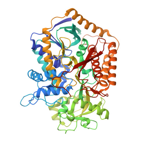
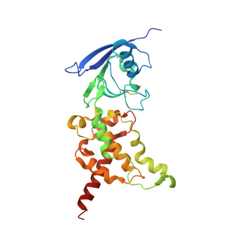
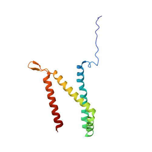
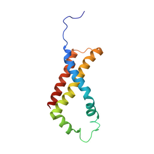
Geometric restraint drives on- and off-pathway catalysis by the Escherichia coli menaquinol:fumarate reductase.

Tomasiak, T.M.Archuleta, T.L.Andrell, J.Luna-Chavez, C.Davis, T.A.Sarwar, M.Ham, A.J.McDonald, W.H.Yankovskaya, V.Stern, H.A.Johnston, J.N.Maklashina, E.Cecchini, G.Iverson, T.M.

(2011) J Biological Chem 286: 3047-3056

  • DOI: https://doi.org/10.1074/jbc.M110.192849
  • Primary Citation Related Structures: 
    3P4P, 3P4Q, 3P4R, 3P4S

  • PubMed Abstract: 

    Complex II superfamily members catalyze the kinetically difficult interconversion of succinate and fumarate. Due to the relative simplicity of complex II substrates and their similarity to other biologically abundant small molecules, substrate specificity presents a challenge in this system. In order to identify determinants for on-pathway catalysis, off-pathway catalysis, and enzyme inhibition, crystal structures of Escherichia coli menaquinol:fumarate reductase (QFR), a complex II superfamily member, were determined bound to the substrate, fumarate, and the inhibitors oxaloacetate, glutarate, and 3-nitropropionate. Optical difference spectroscopy and computational modeling support a model where QFR twists the dicarboxylate, activating it for catalysis. Orientation of the C2-C3 double bond of activated fumarate parallel to the C(4a)-N5 bond of FAD allows orbital overlap between the substrate and the cofactor, priming the substrate for nucleophilic attack. Off-pathway catalysis, such as the conversion of malate to oxaloacetate or the activation of the toxin 3-nitropropionate may occur when inhibitors bind with a similarly activated bond in the same position. Conversely, inhibitors that do not orient an activatable bond in this manner, such as glutarate and citrate, are excluded from catalysis and act as inhibitors of substrate binding. These results support a model where electronic interactions via geometric constraint and orbital steering underlie catalysis by QFR.


  • Organizational Affiliation
    • Department of Pharmacology, Vanderbilt University Medical Center, Nashville, Tennessee 37232, USA.

Macromolecule Content 

  • Total Structure Weight: 240.37 kDa 
  • Atom Count: 16,873 
  • Modeled Residue Count: 2,138 
  • Deposited Residue Count: 2,138 
  • Unique protein chains: 4

Macromolecules

Find similar proteins by:|  3D Structure
Entity ID: 1
MoleculeChains  Sequence LengthOrganismDetailsImage
Fumarate reductase flavoprotein subunitA,
E [auth M]
577Escherichia coli 042Mutation(s): 0 
Gene Names: frdAEC042_4630
EC: 1.3.99.1 (PDB Primary Data), 1.3.5.1 (UniProt)
Membrane Entity: Yes 
UniProt
Find proteins for P00363 (Escherichia coli (strain K12))
Explore P00363 
Go to UniProtKB:  P00363
Entity Groups
Sequence Clusters30% Identity50% Identity70% Identity90% Identity95% Identity100% Identity
UniProt GroupP00363
Sequence Annotations
Expand
Reference Sequence
Find similar proteins by:|  3D Structure
Entity ID: 2
MoleculeChains  Sequence LengthOrganismDetailsImage
Fumarate reductase iron-sulfur proteinB,
F [auth N]
243Escherichia coli 536Mutation(s): 0 
Gene Names: ECP_4399
EC: 1.3.99.1 (PDB Primary Data), 1.3.5.1 (UniProt)
Membrane Entity: Yes 
UniProt
Find proteins for P0AC47 (Escherichia coli (strain K12))
Explore P0AC47 
Go to UniProtKB:  P0AC47
Entity Groups
Sequence Clusters30% Identity50% Identity70% Identity90% Identity95% Identity100% Identity
UniProt GroupP0AC47
Sequence Annotations
Expand
Reference Sequence
Find similar proteins by:|  3D Structure
Entity ID: 3
MoleculeChains  Sequence LengthOrganismDetailsImage
Fumarate reductase subunit CC,
G [auth O]
130Escherichia coli DH1Mutation(s): 0 
Gene Names: frdCEcDH1_3838
Membrane Entity: Yes 
UniProt
Find proteins for P0A8Q0 (Escherichia coli (strain K12))
Explore P0A8Q0 
Go to UniProtKB:  P0A8Q0
Entity Groups
Sequence Clusters30% Identity50% Identity70% Identity90% Identity95% Identity100% Identity
UniProt GroupP0A8Q0
Sequence Annotations
Expand
Reference Sequence
Find similar proteins by:|  3D Structure
Entity ID: 4
MoleculeChains  Sequence LengthOrganismDetailsImage
Fumarate reductase subunit DD,
H [auth P]
119Escherichia coli DH1Mutation(s): 0 
Gene Names: frdDEcDH1_3839
Membrane Entity: Yes 
UniProt
Find proteins for P0A8Q3 (Escherichia coli (strain K12))
Explore P0A8Q3 
Go to UniProtKB:  P0A8Q3
Entity Groups
Sequence Clusters30% Identity50% Identity70% Identity90% Identity95% Identity100% Identity
UniProt GroupP0A8Q3
Sequence Annotations
Expand
Reference Sequence

Small Molecules

Ligands 5 Unique
IDChains Name / Formula / InChI Key2D Diagram3D Interactions
FAD

Query on FAD


Download:Ideal Coordinates CCD File
I [auth A],
N [auth M]
FLAVIN-ADENINE DINUCLEOTIDE
C27 H33 N9 O15 P2
VWWQXMAJTJZDQX-UYBVJOGSSA-N
SF4

Query on SF4


Download:Ideal Coordinates CCD File
M [auth B],
Q [auth N]
IRON/SULFUR CLUSTER
Fe4 S4
LJBDFODJNLIPKO-UHFFFAOYSA-N
F3S

Query on F3S


Download:Ideal Coordinates CCD File
L [auth B],
P [auth N]
FE3-S4 CLUSTER
Fe3 S4
FCXHZBQOKRZXKS-UHFFFAOYSA-N
FES

Query on FES


Download:Ideal Coordinates CCD File
K [auth B],
O [auth N]
FE2/S2 (INORGANIC) CLUSTER
Fe2 S2
NIXDOXVAJZFRNF-UHFFFAOYSA-N
FUM

Query on FUM


Download:Ideal Coordinates CCD File
J [auth A]FUMARIC ACID
C4 H4 O4
VZCYOOQTPOCHFL-OWOJBTEDSA-N

Experimental Data & Validation

Experimental Data

  • Method: X-RAY DIFFRACTION
  • Resolution: 2.80 Å
  • R-Value Free:  0.272 (Depositor), 0.278 (DCC) 
  • R-Value Work:  0.249 (Depositor), 0.258 (DCC) 
  • R-Value Observed: 0.249 (Depositor) 
Space Group: P 21 21 21
Unit Cell:
Length ( Å )Angle ( ˚ )
a = 96.317α = 90
b = 137.633β = 90
c = 270.684γ = 90
Software Package:
Software NamePurpose
REFMACrefinement
PDB_EXTRACTdata extraction
DENZOdata reduction
SCALEPACKdata scaling

Structure Validation

View Full Validation Report


Ligand Structure Quality Assessment 


Entry History 

Revision History  (Full details and data files)

  • Version 1.0: 2010-12-01
    Type: Initial release
  • Version 1.1: 2011-07-13
    Changes: Version format compliance
  • Version 1.2: 2012-02-15
    Changes: Non-polymer description
  • Version 1.3: 2017-11-08
    Changes: Refinement description
  • Version 2.0: 2023-11-15
    Changes: Atomic model, Data collection, Database references, Derived calculations
  • Version 2.1: 2024-10-30
    Changes: Structure summary