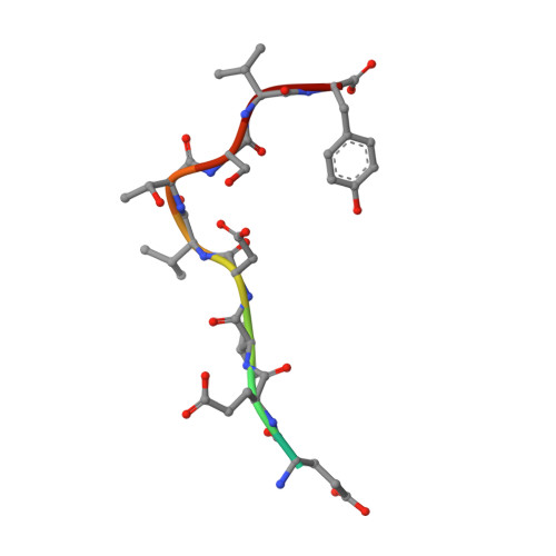
3P4F

Structural and biochemical insights into MLL1 core complex assembly and regulation.


Experimental Data Snapshot

  • Method: X-RAY DIFFRACTION
  • Resolution: 2.35 Å
  • R-Value Free: 0.249 
  • R-Value Work: 0.194 
  • R-Value Observed: 0.197 

Starting Model: experimental
View more details

wwPDB Validation   3D Report Full Report


This is version 1.4 of the entry. See complete history


Literature

Structural and biochemical insights into MLL1 core complex assembly.

Avdic, V.Zhang, P.Lanouette, S.Groulx, A.Tremblay, V.Brunzelle, J.Couture, J.-F.

(2011) Structure 19: 101-108

  • DOI: https://doi.org/10.1016/j.str.2010.09.022
  • Primary Citation of Related Structures:  
    3P4F

  • PubMed Abstract: 

    Histone H3 Lys-4 methylation is predominantly catalyzed by a family of methyltransferases whose enzymatic activity depends on their interaction with a three-subunit complex composed of WDR5, RbBP5, and Ash2L. Here, we report that a segment of 50 residues of RbBP5 bridges the Ash2L C-terminal domain to WDR5. The crystal structure of WDR5 in ternary complex with RbBP5 and MLL1 reveals that both proteins binds peptide-binding clefts located on opposite sides of WDR5's β-propeller domain. RbBP5 engages in several hydrogen bonds and van der Waals contacts within a V-shaped cleft formed by the junction of two blades on WDR5. Mutational analyses of both the WDR5 V-shaped cleft and RbBP5 residues reveal that the interactions between RbBP5 and WDR5 are important for the stimulation of MLL1 methyltransferase activity. Overall, this study provides the structural basis underlying the formation of the WDR5-RbBP5 subcomplex and further highlight the crucial role of WDR5 in scaffolding the MLL1 core complex.


  • Organizational Affiliation

    Ottawa Institute of Systems Biology, Department of Biochemistry, Microbiology and Immunology, University of Ottawa, Ottawa, ON K1H 8M5, Canada.


Macromolecules
Find similar proteins by:  (by identity cutoff)  |  3D Structure
Entity ID: 1
MoleculeChains Sequence LengthOrganismDetailsImage
WD repeat-containing protein 5317Homo sapiensMutation(s): 0 
Gene Names: WDR5BIG3
UniProt & NIH Common Fund Data Resources
Find proteins for P61964 (Homo sapiens)
Explore P61964 
Go to UniProtKB:  P61964
PHAROS:  P61964
GTEx:  ENSG00000196363 
Entity Groups  
Sequence Clusters30% Identity50% Identity70% Identity90% Identity95% Identity100% Identity
UniProt GroupP61964
Sequence Annotations
Expand
  • Reference Sequence

Find similar proteins by:  Sequence   |   3D Structure  

Entity ID: 2
MoleculeChains Sequence LengthOrganismDetailsImage
Retinoblastoma-binding protein 511Homo sapiensMutation(s): 1 
UniProt & NIH Common Fund Data Resources
Find proteins for Q15291 (Homo sapiens)
Explore Q15291 
Go to UniProtKB:  Q15291
PHAROS:  Q15291
GTEx:  ENSG00000117222 
Entity Groups  
Sequence Clusters30% Identity50% Identity70% Identity90% Identity95% Identity100% Identity
UniProt GroupQ15291
Sequence Annotations
Expand
  • Reference Sequence

Find similar proteins by:  Sequence   |   3D Structure  

Entity ID: 3
MoleculeChains Sequence LengthOrganismDetailsImage
Histone-lysine N-methyltransferase MLL10Homo sapiensMutation(s): 1 
EC: 2.1.1.43
UniProt & NIH Common Fund Data Resources
Find proteins for Q03164 (Homo sapiens)
Explore Q03164 
Go to UniProtKB:  Q03164
PHAROS:  Q03164
GTEx:  ENSG00000118058 
Entity Groups  
Sequence Clusters30% Identity50% Identity70% Identity90% Identity95% Identity100% Identity
UniProt GroupQ03164
Sequence Annotations
Expand
  • Reference Sequence
Experimental Data & Validation

Experimental Data

  • Method: X-RAY DIFFRACTION
  • Resolution: 2.35 Å
  • R-Value Free: 0.249 
  • R-Value Work: 0.194 
  • R-Value Observed: 0.197 
  • Space Group: P 21 21 21
Unit Cell:
Length ( Å )Angle ( ˚ )
a = 53.85α = 90
b = 81.35β = 90
c = 81.78γ = 90
Software Package:
Software NamePurpose
StructureStudiodata collection
PHASERphasing
BUSTERrefinement
d*TREKdata reduction
d*TREKdata scaling

Structure Validation

View Full Validation Report


Entry History 

Deposition Data

Revision History  (Full details and data files)

  • Version 1.0: 2010-12-08
    Type: Initial release
  • Version 1.1: 2011-07-13
    Changes: Version format compliance
  • Version 1.2: 2014-10-08
    Changes: Database references
  • Version 1.3: 2018-02-07
    Changes: Experimental preparation
  • Version 1.4: 2023-09-06
    Changes: Data collection, Database references, Refinement description