


Crystal structure of isocitrate lyase from Brucella melitensis, bound to magnesium isocitrate
SSGCID, Gardberg, A., Edwards, T., Gander, M., Staker, B., Stewart, L.To be published.
Experimental Data Snapshot
Starting Model: experimental
View more details
Entity ID: 1 | |||||
|---|---|---|---|---|---|
| Molecule | Chains | Sequence Length | Organism | Details | Image |
| Isocitrate lyase | 433 | Brucella abortus 2308 | Mutation(s): 0 Gene Names: aceA, BAB1_1631, BruAb1_1601 EC: 4.1.3.1 | 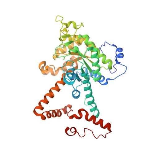 | |
UniProt | |||||
Entity Groups | |||||
| Sequence Clusters | 30% Identity50% Identity70% Identity90% Identity95% Identity100% Identity | ||||
| UniProt Group | Q2YQA0 | ||||
Sequence AnnotationsExpand | |||||
Reference Sequence | |||||
| Ligands 3 Unique | |||||
|---|---|---|---|---|---|
| ID | Chains | Name / Formula / InChI Key | 2D Diagram | 3D Interactions | |
| ICT Download:Ideal Coordinates CCD File | G [auth A], J [auth B], N [auth C], Q [auth D] | ISOCITRIC ACID C6 H8 O7 ODBLHEXUDAPZAU-ZAFYKAAXSA-N |  | ||
| EDO Download:Ideal Coordinates CCD File | E [auth A] F [auth A] I [auth B] L [auth C] M [auth C] | 1,2-ETHANEDIOL C2 H6 O2 LYCAIKOWRPUZTN-UHFFFAOYSA-N |  | ||
| MG Download:Ideal Coordinates CCD File | H [auth A], K [auth B], O [auth C], R [auth D] | MAGNESIUM ION Mg JLVVSXFLKOJNIY-UHFFFAOYSA-N |  | ||
| Length ( Å ) | Angle ( ˚ ) |
|---|---|
| a = 76.91 | α = 90 |
| b = 135.93 | β = 90 |
| c = 181.79 | γ = 90 |
| Software Name | Purpose |
|---|---|
| XSCALE | data scaling |
| PHASER | phasing |
| REFMAC | refinement |
| PDB_EXTRACT | data extraction |
| StructureStudio | data collection |
| XDS | data reduction |


