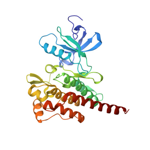
3OY3

Crystal structure of ABL T315I mutant kinase domain bound with a DFG-out inhibitor AP24589


Experimental Data Snapshot

  • Method: X-RAY DIFFRACTION
  • Resolution: 1.95 Å
  • R-Value Free: 0.266 
  • R-Value Work: 0.220 

Starting Model: experimental
View more details

wwPDB Validation   3D Report Full Report


Ligand Structure Quality Assessment 


This is version 1.2 of the entry. See complete history


Literature

Structural Mechanism of the Pan-BCR-ABL Inhibitor Ponatinib (AP24534): Lessons for Overcoming Kinase Inhibitor Resistance.

Zhou, T.Commodore, L.Huang, W.S.Wang, Y.Thomas, M.Keats, J.Xu, Q.Rivera, V.M.Shakespeare, W.C.Clackson, T.Dalgarno, D.C.Zhu, X.

(2011) Chem Biol Drug Des 77: 1-11

  • DOI: https://doi.org/10.1111/j.1747-0285.2010.01054.x
  • Primary Citation of Related Structures:  
    3OXZ, 3OY3

  • PubMed Abstract: 

    The BCR-ABL inhibitor imatinib has revolutionized the treatment of chronic myeloid leukemia. However, drug resistance caused by kinase domain mutations has necessitated the development of new mutation-resistant inhibitors, most recently against the T315I gatekeeper residue mutation. Ponatinib (AP24534) inhibits both native and mutant BCR-ABL, including T315I, acting as a pan-BCR-ABL inhibitor. Here, we undertook a combined crystallographic and structure-activity relationship analysis on ponatinib to understand this unique profile. While the ethynyl linker is a key inhibitor functionality that interacts with the gatekeeper, virtually all other components of ponatinib play an essential role in its T315I inhibitory activity. The extensive network of optimized molecular contacts found in the DFG-out binding mode leads to high potency and renders binding less susceptible to disruption by single point mutations. The inhibitory mechanism exemplified by ponatinib may have broad relevance to designing inhibitors against other kinases with mutated gatekeeper residues.


  • Organizational Affiliation

    ARIAD Pharmaceuticals Inc., 26 Landsdowne Street, Cambridge, MA 02139, USA.


Macromolecules
Find similar proteins by:  (by identity cutoff)  |  3D Structure
Entity ID: 1
MoleculeChains Sequence LengthOrganismDetailsImage
Tyrosine-protein kinase ABL1
A, B
284Mus musculusMutation(s): 1 
Gene Names: Abl1Abl
EC: 2.7.10.2
UniProt
Find proteins for P00520 (Mus musculus)
Explore P00520 
Go to UniProtKB:  P00520
Entity Groups  
Sequence Clusters30% Identity50% Identity70% Identity90% Identity95% Identity100% Identity
UniProt GroupP00520
Sequence Annotations
Expand
  • Reference Sequence
Small Molecules
Ligands 1 Unique
IDChains Name / Formula / InChI Key2D Diagram3D Interactions
XY3
Query on XY3

Download Ideal Coordinates CCD File 
C [auth A],
D [auth B]
5-[(5-{[4-{[4-(2-hydroxyethyl)piperazin-1-yl]methyl}-3-(trifluoromethyl)phenyl]carbamoyl}-2-methylphenyl)ethynyl]-1-methyl-1H-imidazole-2-carboxamide
C29 H31 F3 N6 O3
GOLRXYBFRBKBAG-UHFFFAOYSA-N
Binding Affinity Annotations 
IDSourceBinding Affinity
XY3 BindingDB:  3OY3 IC50: 3.6 (nM) from 1 assay(s)
PDBBind:  3OY3 IC50: 35.8 (nM) from 1 assay(s)
Experimental Data & Validation

Experimental Data

  • Method: X-RAY DIFFRACTION
  • Resolution: 1.95 Å
  • R-Value Free: 0.266 
  • R-Value Work: 0.220 
  • Space Group: P 1 21 1
Unit Cell:
Length ( Å )Angle ( ˚ )
a = 66.471α = 90
b = 59.988β = 97.97
c = 89.439γ = 90
Software Package:
Software NamePurpose
CNSrefinement
PDB_EXTRACTdata extraction
HKL-2000data collection
HKL-2000data reduction
HKL-2000data scaling
AMoREphasing

Structure Validation

View Full Validation Report


Ligand Structure Quality Assessment 


Entry History 

Deposition Data

Revision History  (Full details and data files)

  • Version 1.0: 2010-12-15
    Type: Initial release
  • Version 1.1: 2011-07-13
    Changes: Version format compliance
  • Version 1.2: 2023-09-06
    Changes: Data collection, Database references, Derived calculations, Refinement description