3OX5

Crystal Structure of the calcium sensor calcium-binding protein 1 (CaBP1)


Experimental Data Snapshot

  • Method: X-RAY DIFFRACTION
  • Resolution: 2.90 Å
  • R-Value Free: 0.250 
  • R-Value Work: 0.213 
  • R-Value Observed: 0.215 

wwPDB Validation   3D Report Full Report


This is version 1.2 of the entry. See complete history


Literature

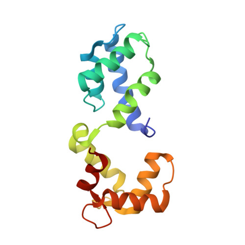
Structural Basis for the Differential Effects of CaBP1 and Calmodulin on Ca(V)1.2 Calcium-Dependent Inactivation.

Findeisen, F.Minor, D.L.

(2010) Structure 18: 1617-1631

  • DOI: https://doi.org/10.1016/j.str.2010.09.012
  • Primary Citation of Related Structures:  
    3OX5, 3OX6

  • PubMed Abstract: 

    Calcium-binding protein 1 (CaBP1), a calmodulin (CaM) homolog, endows certain voltage-gated calcium channels (Ca(V)s) with unusual properties. CaBP1 inhibits Ca(V)1.2 calcium-dependent inactivation (CDI) and introduces calcium-dependent facilitation (CDF). Here, we show that the ability of CaBP1 to inhibit Ca(V)1.2 CDI and induce CDF arises from interaction between the CaBP1 N-lobe and interlobe linker residue Glu94. Unlike CaM, where functional EF hands are essential for channel modulation, CDI inhibition does not require functional CaBP1 EF hands. Furthermore, CaBP1-mediated CDF has different molecular requirements than CaM-mediated CDF. Overall, the data show that CaBP1 comprises two structural modules having separate functions: similar to CaM, the CaBP1 C-lobe serves as a high-affinity anchor that binds the Ca(V)1.2 IQ domain at a site that overlaps with the Ca²+/CaM C-lobe site, whereas the N-lobe/linker module houses the elements required for channel modulation. Discovery of this division provides the framework for understanding how CaBP1 regulates Ca(V)s.


  • Organizational Affiliation

    Cardiovascular Research Institute, University of California, San Francisco, CA 94158-2330, USA.


Macromolecules
Find similar proteins by:  (by identity cutoff)  |  3D Structure
Entity ID: 1
MoleculeChains Sequence LengthOrganismDetailsImage
Calcium-binding protein 1
A, B, C, D, E
A, B, C, D, E, F
153Homo sapiensMutation(s): 0 
Gene Names: CABP1
UniProt & NIH Common Fund Data Resources
Find proteins for Q9NZU7 (Homo sapiens)
Explore Q9NZU7 
Go to UniProtKB:  Q9NZU7
PHAROS:  Q9NZU7
GTEx:  ENSG00000157782 
Entity Groups  
Sequence Clusters30% Identity50% Identity70% Identity90% Identity95% Identity100% Identity
UniProt GroupQ9NZU7
Sequence Annotations
Expand
  • Reference Sequence
Small Molecules
Ligands 1 Unique
IDChains Name / Formula / InChI Key2D Diagram3D Interactions
CA
Query on CA

Download Ideal Coordinates CCD File 
G [auth A]
H [auth A]
I [auth B]
J [auth B]
K [auth C]
G [auth A],
H [auth A],
I [auth B],
J [auth B],
K [auth C],
L [auth C],
M [auth D],
N [auth D],
O [auth E],
P [auth E],
Q [auth F],
R [auth F]
CALCIUM ION
Ca
BHPQYMZQTOCNFJ-UHFFFAOYSA-N
Experimental Data & Validation

Experimental Data

  • Method: X-RAY DIFFRACTION
  • Resolution: 2.90 Å
  • R-Value Free: 0.250 
  • R-Value Work: 0.213 
  • R-Value Observed: 0.215 
  • Space Group: I 2 3
Unit Cell:
Length ( Å )Angle ( ˚ )
a = 206.044α = 90
b = 206.044β = 90
c = 206.044γ = 90
Software Package:
Software NamePurpose
HKL-2000data collection
PHENIXmodel building
REFMACrefinement
MOSFLMdata reduction
SCALAdata scaling
PHENIXphasing

Structure Validation

View Full Validation Report


Entry History 

Deposition Data

Revision History  (Full details and data files)

  • Version 1.0: 2010-12-22
    Type: Initial release
  • Version 1.1: 2011-07-13
    Changes: Version format compliance
  • Version 1.2: 2024-02-21
    Changes: Data collection, Database references, Derived calculations