3OO9

Crystal structures and biochemical characterization of the bacterial solute receptor AcbH reveal an unprecedented exclusive substrate preference for b-D-galactopyranose


Experimental Data Snapshot

  • Method: X-RAY DIFFRACTION
  • Resolution: 1.76 Å
  • R-Value Free: 0.195 
  • R-Value Work: 0.155 
  • R-Value Observed: 0.157 

wwPDB Validation   3D Report Full Report


This is version 1.4 of the entry. See complete history


Literature

Crystal structures of the bacterial solute receptor AcbH displaying an exclusive substrate preference for beta-D-galactopyranose

Licht, A.Bulut, H.Scheffel, F.Daumke, O.Wehmeier, U.F.Saenger, W.Schneider, E.Vahedi-Faridi, A.

(2011) J Mol Biol 406: 92-105

  • DOI: https://doi.org/10.1016/j.jmb.2010.11.048
  • Primary Citation of Related Structures:  
    3OO6, 3OO7, 3OO8, 3OO9, 3OOA

  • PubMed Abstract: 

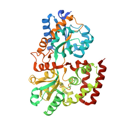
    Solute receptors (binding proteins) are indispensable components of canonical ATP-binding cassette importers in prokaryotes. Here, we report on the characterization and crystal structures in the closed and open conformations of AcbH, the solute receptor of the putative carbohydrate transporter AcbFG which is encoded in the acarbose (acarviosyl-1,4-maltose) biosynthetic gene cluster from Actinoplanes sp. SE50/110. Binding assays identified AcbH as a high-affinity monosaccharide-binding protein with a dissociation constant (K(d)) for β-d-galactopyranose of 9.8±1.0 nM. Neither galactose-containing di- and trisaccharides, such as lactose and raffinose, nor monosaccharides including d-galacturonic acid, l-arabinose, d-xylose and l-rhamnose competed with [(1)(4)C]galactose for binding to AcbH. Moreover, AcbH does not bind d-glucose, which is a common property of all but one d-galactose-binding proteins characterized to date. Strikingly, determination of the X-ray structure revealed that AcbH is structurally homologous to maltose-binding proteins rather than to glucose-binding proteins. Two helices are inserted in the substrate-binding pocket, which reduces the cavity size and allows the exclusive binding of monosaccharides, specifically β-d-galactopyranose, in the (4)C(1) conformation. Site-directed mutagenesis of three residues from the binding pocket (Arg82, Asp361 and Arg362) that interact with the axially oriented O4-H hydroxyl of the bound galactopyranose and subsequent functional analysis indicated that these residues are crucial for galactose binding. To our knowledge, this is the first report of the tertiary structure of a solute receptor with exclusive affinity for β-d-galactopyranose. The putative role of a galactose import system in the context of acarbose metabolism in Actinoplanes sp. is discussed.


  • Organizational Affiliation

    Institut für Biologie, AG Bakterienphysiologie, Humboldt Universität zu Berlin, Chausseestr. 117, D-10115 Berlin, Germany.


Macromolecules
Find similar proteins by:  (by identity cutoff)  |  3D Structure
Entity ID: 1
MoleculeChains Sequence LengthOrganismDetailsImage
ABC transporter binding protein AcbH
A, B
415Actinoplanes sp. SE50/110Mutation(s): 2 
Gene Names: AcbH
UniProt
Find proteins for Q27GR2 (Actinoplanes sp. (strain ATCC 31044 / CBS 674.73 / SE50/110))
Explore Q27GR2 
Go to UniProtKB:  Q27GR2
Entity Groups  
Sequence Clusters30% Identity50% Identity70% Identity90% Identity95% Identity100% Identity
UniProt GroupQ27GR2
Sequence Annotations
Expand
  • Reference Sequence
Small Molecules
Ligands 2 Unique
IDChains Name / Formula / InChI Key2D Diagram3D Interactions
SO4
Query on SO4

Download Ideal Coordinates CCD File 
C [auth A]
D [auth A]
E [auth A]
F [auth A]
G [auth A]
C [auth A],
D [auth A],
E [auth A],
F [auth A],
G [auth A],
H [auth A],
I [auth A],
J [auth A],
K [auth A],
N [auth B],
O [auth B],
P [auth B],
Q [auth B],
R [auth B],
S [auth B],
T [auth B],
U [auth B]
SULFATE ION
O4 S
QAOWNCQODCNURD-UHFFFAOYSA-L
GOL
Query on GOL

Download Ideal Coordinates CCD File 
AA [auth B]
L [auth A]
M [auth A]
V [auth B]
W [auth B]
AA [auth B],
L [auth A],
M [auth A],
V [auth B],
W [auth B],
X [auth B],
Y [auth B],
Z [auth B]
GLYCEROL
C3 H8 O3
PEDCQBHIVMGVHV-UHFFFAOYSA-N
Experimental Data & Validation

Experimental Data

  • Method: X-RAY DIFFRACTION
  • Resolution: 1.76 Å
  • R-Value Free: 0.195 
  • R-Value Work: 0.155 
  • R-Value Observed: 0.157 
  • Space Group: P 1 21 1
Unit Cell:
Length ( Å )Angle ( ˚ )
a = 39.25α = 90
b = 65.76β = 90.03
c = 177.72γ = 90
Software Package:
Software NamePurpose
XSCALEdata scaling
REFMACrefinement
PDB_EXTRACTdata extraction
ADSCdata collection
PHASERphasing

Structure Validation

View Full Validation Report


Entry History 

Deposition Data

Revision History  (Full details and data files)

  • Version 1.0: 2010-12-29
    Type: Initial release
  • Version 1.1: 2011-07-13
    Changes: Version format compliance
  • Version 1.2: 2014-03-26
    Changes: Database references
  • Version 1.3: 2024-03-20
    Changes: Data collection, Database references, Derived calculations
  • Version 1.4: 2024-04-03
    Changes: Refinement description