3OO4

R-state human hemoglobin: nitriheme modified at alpha


Experimental Data Snapshot

  • Method: X-RAY DIFFRACTION
  • Resolution: 1.90 Å
  • R-Value Free: 0.263 
  • R-Value Work: 0.207 
  • R-Value Observed: 0.210 

wwPDB Validation   3D Report Full Report


Ligand Structure Quality Assessment 


This is version 1.2 of the entry. See complete history


Literature

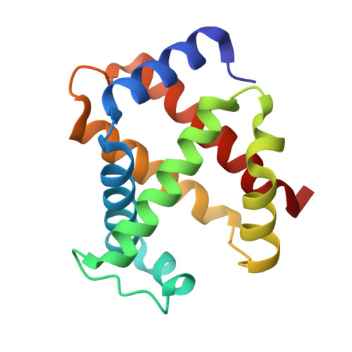
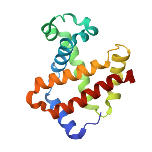
Crystallographic Trapping of Heme Loss Intermediates during the Nitrite-Induced Degradation of Human Hemoglobin.

Yi, J.Thomas, L.M.Musayev, F.N.Safo, M.K.Richter-Addo, G.B.

(2011) Biochemistry 50: 8323-8332

  • DOI: https://doi.org/10.1021/bi2009322
  • Primary Citation of Related Structures:  
    3ONZ, 3OO4, 3OO5

  • PubMed Abstract: 

    Heme is an important cofactor in a large number of essential proteins and is often involved in small molecule binding and activation. Loss of heme from proteins thus negatively affects the function of these proteins but is also an important component of iron recycling. The characterization of intermediates that form during the loss of heme from proteins has been problematic, in a large part, because of the instability of such intermediates. We have characterized, by X-ray crystallography, three compounds that form during the nitrite-induced degradation of human α(2)β(2) hemoglobin (Hb). The first is an unprecedented complex that exhibits a large β heme displacement of 4.8 Å toward the protein exterior; the heme displacement is stabilized by the binding of the distal His residue to the heme Fe, which in turn allows for the unusual binding of an exogenous ligand on the proximal face of the heme. We have also structurally characterized complexes that display regiospecific nitration of the heme at the 2-vinyl position; we show that heme nitration is not a prerequisite for heme loss. Our results provide structural insight into a possible pathway for nitrite-induced loss of heme from human Hb.


  • Organizational Affiliation

    Department of Chemistry and Biochemistry, University of Oklahoma, Norman, Oklahoma 73019, United States. yijun@ou.edu


Macromolecules
Find similar proteins by:  (by identity cutoff)  |  3D Structure
Entity ID: 1
MoleculeChains Sequence LengthOrganismDetailsImage
Hemoglobin subunit alpha141Homo sapiensMutation(s): 0 
UniProt & NIH Common Fund Data Resources
Find proteins for P69905 (Homo sapiens)
Explore P69905 
Go to UniProtKB:  P69905
PHAROS:  P69905
Entity Groups  
Sequence Clusters30% Identity50% Identity70% Identity90% Identity95% Identity100% Identity
UniProt GroupP69905
Sequence Annotations
Expand
  • Reference Sequence
Find similar proteins by:  (by identity cutoff)  |  3D Structure
Entity ID: 2
MoleculeChains Sequence LengthOrganismDetailsImage
Hemoglobin subunit beta146Homo sapiensMutation(s): 0 
UniProt & NIH Common Fund Data Resources
Find proteins for P68871 (Homo sapiens)
Explore P68871 
Go to UniProtKB:  P68871
PHAROS:  P68871
GTEx:  ENSG00000244734 
Entity Groups  
Sequence Clusters30% Identity50% Identity70% Identity90% Identity95% Identity100% Identity
UniProt GroupP68871
Sequence Annotations
Expand
  • Reference Sequence
Small Molecules
Ligands 3 Unique
IDChains Name / Formula / InChI Key2D Diagram3D Interactions
NTE
Query on NTE

Download Ideal Coordinates CCD File 
C [auth A][3,3'-{7-ethenyl-3,8,13,17-tetramethyl-12-[(E)-2-nitroethenyl]porphyrin-2,18-diyl-kappa~4~N~21~,N~22~,N~23~,N~24~}dipro panoato(2-)]iron
C34 H31 Fe N5 O6
MBEUICWDKPMPCN-RXJYIKGQSA-L
HEM
Query on HEM

Download Ideal Coordinates CCD File 
E [auth B]PROTOPORPHYRIN IX CONTAINING FE
C34 H32 Fe N4 O4
KABFMIBPWCXCRK-RGGAHWMASA-L
NO2
Query on NO2

Download Ideal Coordinates CCD File 
D [auth A],
F [auth B]
NITRITE ION
N O2
IOVCWXUNBOPUCH-UHFFFAOYSA-M
Experimental Data & Validation

Experimental Data

  • Method: X-RAY DIFFRACTION
  • Resolution: 1.90 Å
  • R-Value Free: 0.263 
  • R-Value Work: 0.207 
  • R-Value Observed: 0.210 
  • Space Group: P 41 21 2
Unit Cell:
Length ( Å )Angle ( ˚ )
a = 53.487α = 90
b = 53.487β = 90
c = 190.001γ = 90
Software Package:
Software NamePurpose
CrystalCleardata collection
PHASERphasing
PHENIXrefinement
d*TREKdata reduction
d*TREKdata scaling

Structure Validation

View Full Validation Report


Ligand Structure Quality Assessment 


Entry History 

Deposition Data

Revision History  (Full details and data files)

  • Version 1.0: 2011-09-07
    Type: Initial release
  • Version 1.1: 2011-10-12
    Changes: Database references
  • Version 1.2: 2023-09-06
    Changes: Data collection, Database references, Derived calculations, Refinement description, Structure summary