Crystal structure of UBA2(ufd)-Ubc9: insights into E1-E2 interactions in Sumo pathways.
Wang, J., Taherbhoy, A.M., Hunt, H.W., Seyedin, S.N., Miller, D.W., Miller, D.J., Huang, D.T., Schulman, B.A.(2010) PLoS One 5: e15805-e15805
- PubMed: 21209884 Search on PubMedSearch on PubMed Central
- DOI: https://doi.org/10.1371/journal.pone.0015805
- Primary Citation Related Structures:
3ONG, 3ONH - PubMed Abstract:
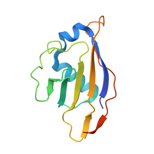
Canonical ubiquitin-like proteins (UBLs) such as ubiquitin, Sumo, NEDD8, and ISG15 are ligated to targets by E1-E2-E3 multienzyme cascades. The Sumo cascade, conserved among all eukaryotes, regulates numerous biological processes including protein localization, transcription, DNA replication, and mitosis. Sumo conjugation is initiated by the heterodimeric Aos1-Uba2 E1 enzyme (in humans called Sae1-Uba2), which activates Sumo's C-terminus, binds the dedicated E2 enzyme Ubc9, and promotes Sumo C-terminal transfer between the Uba2 and Ubc9 catalytic cysteines. To gain insights into details of E1-E2 interactions in the Sumo pathway, we determined crystal structures of the C-terminal ubiquitin fold domain (ufd) from yeast Uba2 (Uba2(ufd)), alone and in complex with Ubc9. The overall structures of both yeast Uba2(ufd) and Ubc9 superimpose well on their individual human counterparts, suggesting conservation of fundamental features of Sumo conjugation. Docking the Uba2(ufd)-Ubc9 and prior full-length human Uba2 structures allows generation of models for steps in Sumo transfer from Uba2 to Ubc9, and supports the notion that Uba2 undergoes remarkable conformational changes during the reaction. Comparisons to previous structures from the NEDD8 cascade demonstrate that UBL cascades generally utilize some parallel E1-E2 interaction surfaces. In addition, the structure of the Uba2(ufd)-Ubc9 complex reveals interactions unique to Sumo E1 and E2. Comparison with a previous Ubc9-E3 complex structure demonstrates overlap between Uba2 and E3 binding sites on Ubc9, indicating that loading with Sumo and E3-catalyzed transfer to substrates are strictly separate steps. The results suggest mechanisms establishing specificity and order in Sumo conjugation cascades.
- Department of Structural Biology, St. Jude Children's Research Hospital, Memphis, Tennessee, United States of America.
Organizational Affiliation: