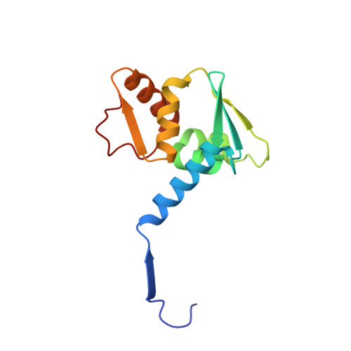
The structure of the Bach2 POZ-domain dimer reveals an intersubunit disulfide bond.
Rosbrook, G.O., Stead, M.A., Carr, S.B., Wright, S.C.(2012) Acta Crystallogr D Biol Crystallogr 68: 26-34
- PubMed: 22194330 Search on PubMed
- DOI: https://doi.org/10.1107/S0907444911048335
- Primary Citation Related Structures:
3OHU, 3OHV - PubMed Abstract:
Bach2 is a transcriptional repressor that is expressed during specific stages of B-cell development and in neuronal cells. It plays a critical role in modulating class-switch recombination during the differentiation of mature B cells to antibody-secreting plasma cells and it is also an important regulator of apoptotic responses to oxidative stress. Bach2 has been implicated both as an oncogene and as a tumour suppressor in human malignancy. The interaction of Bach2 with its target genes is mediated via its basic leucine-zipper region, whereas the N-terminal POZ domain recruits transcriptional co-repressors and class II histone deacetylases. Here, the crystal structure of the human Bach2 POZ domain is reported at 2.1 Å resolution. The Bach2 POZ-domain dimer resembles the POZ-domain dimers of the POZ zinc finger transcription factors and dimerization is independent of an N-terminal region that has previously been implicated in the dimerization of the POZ basic leucine-zipper protein Bach1. The Bach2 POZ domain crystallized in two forms which differed by the presence of an intersubunit disulfide bond. The intersubunit disulfide bond is present both in bacterially expressed Bach2 POZ domain in solution and in protein expressed in transfected eukaryotic cells. These crystal structures will be relevant for understanding the regulation of Bach2 in response to oxidative stress and for the design of therapeutics that target the Bach2 POZ domain in human malignancy.
- Faculty of Biological Sciences, University of Leeds, Leeds, England.
Organizational Affiliation: