3ODI

Crystal structure of cyclophilin A in complex with Voclosporin E-ISA247


Experimental Data Snapshot

  • Method: X-RAY DIFFRACTION
  • Resolution: 2.20 Å
  • R-Value Free: 0.220 
  • R-Value Work: 0.164 
  • R-Value Observed: 0.166 

wwPDB Validation   3D Report Full Report


This is version 1.4 of the entry. See complete history


Literature

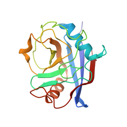
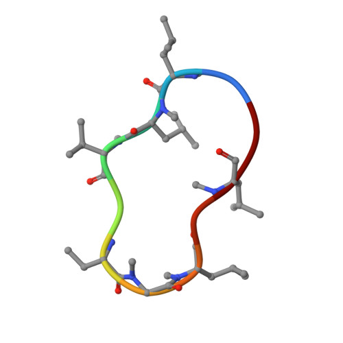
Structural basis for the cyclophilin A binding affinity and immunosuppressive potency of E-ISA247 (voclosporin).

Kuglstatter, A.Mueller, F.Kusznir, E.Gsell, B.Stihle, M.Thoma, R.Benz, J.Aspeslet, L.Freitag, D.Hennig, M.

(2011) Acta Crystallogr D Biol Crystallogr 67: 119-123

  • DOI: https://doi.org/10.1107/S0907444910051905
  • Primary Citation of Related Structures:  
    3ODI, 3ODL

  • PubMed Abstract: 

    E-ISA247 (voclosporin) is a cyclosporin A analogue that is in late-stage clinical development for the treatment of autoimmune diseases and the prevention of organ graft rejection. The X-ray crystal structures of E-ISA247 and its stereoisomer Z-ISA247 bound to cyclophilin A have been determined and their binding affinities were measured to be 15 and 61 nM, respectively, by fluorescence spectroscopy. The higher affinity of E-ISA247 can be explained by superior van der Waals contacts between its unique side chain and cyclophilin A. Comparison with the known ternary structure including calcineurin suggests that the higher immunosuppressive efficacy of E-ISA247 relative to cyclosporin A could be a consequence of structural changes in calcineurin induced by the modified E-ISA247 side chain.


  • Organizational Affiliation

    F. Hoffmann-La Roche, Discovery Technologies, 4070 Basel, Switzerland.


Macromolecules
Find similar proteins by:  (by identity cutoff)  |  3D Structure
Entity ID: 1
MoleculeChains Sequence LengthOrganismDetailsImage
Cyclophilin A
A, C, E, G, I
A, C, E, G, I, K, M, O, Q, S
165Homo sapiensMutation(s): 0 
Gene Names: PPIA
EC: 5.2.1.8
UniProt & NIH Common Fund Data Resources
Find proteins for P62937 (Homo sapiens)
Explore P62937 
Go to UniProtKB:  P62937
PHAROS:  P62937
GTEx:  ENSG00000196262 
Entity Groups  
Sequence Clusters30% Identity50% Identity70% Identity90% Identity95% Identity100% Identity
UniProt GroupP62937
Sequence Annotations
Expand
  • Reference Sequence

Find similar proteins by:  Sequence   |   3D Structure  

Entity ID: 2
MoleculeChains Sequence LengthOrganismDetailsImage
Voclosporin
B, D, F, H, J
B, D, F, H, J, L, N, P, R, T
11Tolypocladium inflatumMutation(s): 1 
Entity Groups  
Sequence Clusters30% Identity50% Identity70% Identity90% Identity95% Identity100% Identity
Sequence Annotations
Expand
  • Reference Sequence
Small Molecules
Modified Residues  4 Unique
IDChains TypeFormula2D DiagramParent
ABA
Query on ABA
B, D, F, H, J
B, D, F, H, J, L, N, P, R, T
L-PEPTIDE LINKINGC4 H9 N O2ALA
MLE
Query on MLE
B, D, F, H, J
B, D, F, H, J, L, N, P, R, T
L-PEPTIDE LINKINGC7 H15 N O2LEU
MVA
Query on MVA
B, D, F, H, J
B, D, F, H, J, L, N, P, R, T
L-PEPTIDE LINKINGC6 H13 N O2VAL
SAR
Query on SAR
B, D, F, H, J
B, D, F, H, J, L, N, P, R, T
PEPTIDE LINKINGC3 H7 N O2GLY
Experimental Data & Validation

Experimental Data

  • Method: X-RAY DIFFRACTION
  • Resolution: 2.20 Å
  • R-Value Free: 0.220 
  • R-Value Work: 0.164 
  • R-Value Observed: 0.166 
  • Space Group: P 1 21 1
Unit Cell:
Length ( Å )Angle ( ˚ )
a = 69.179α = 90
b = 161.341β = 100.06
c = 93.654γ = 90
Software Package:
Software NamePurpose
MAR345dtbdata collection
MOLREPphasing
REFMACrefinement
DENZOdata reduction
SCALEPACKdata scaling

Structure Validation

View Full Validation Report


Entry History 

Deposition Data

Revision History  (Full details and data files)

  • Version 1.0: 2011-02-16
    Type: Initial release
  • Version 1.1: 2011-07-13
    Changes: Version format compliance
  • Version 1.2: 2021-10-06
    Changes: Database references, Derived calculations
  • Version 1.3: 2023-09-06
    Changes: Data collection, Refinement description
  • Version 1.4: 2023-12-06
    Changes: Data collection