3OC9 | pdb_00003oc9

Crystal structure of putative UDP-N-acetylglucosamine pyrophosphorylase from Entamoeba histolytica


Experimental Data Snapshot

  • Method: X-RAY DIFFRACTION
  • Resolution: 1.80 Å
  • R-Value Free: 
    0.227 (Depositor), 0.232 (DCC) 
  • R-Value Work: 
    0.189 (Depositor), 0.190 (DCC) 
  • R-Value Observed: 
    0.191 (Depositor) 

Starting Model: experimental
View more details

wwPDB Validation 3D Report Full Report

Validation slider image for 3OC9

This is version 1.4 of the entry. See complete history

Literature

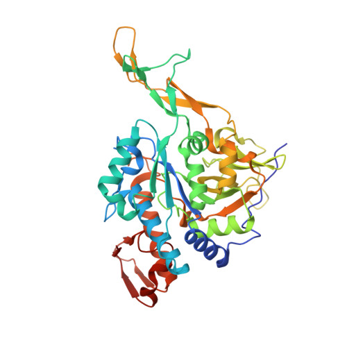
Structure of uridine diphosphate N-acetylglucosamine pyrophosphorylase from Entamoeba histolytica.

Edwards, T.E.Gardberg, A.S.Phan, I.Q.Zhang, Y.Staker, B.L.Myler, P.J.Lorimer, D.D.

(2015) Acta Crystallogr Sect F Struct Biol Cryst Commun 71: 560-565

  • DOI: https://doi.org/10.1107/S2053230X1500179X
  • Primary Citation Related Structures: 
    3OC9

  • PubMed Abstract: 

    Uridine diphosphate N-acetylglucosamine pyrophosphorylase (UAP) catalyzes the final step in the synthesis of UDP-GlcNAc, which is involved in cell-wall biogenesis in plants and fungi and in protein glycosylation. Small-molecule inhibitors have been developed against UAP from Trypanosoma brucei that target an allosteric pocket to provide selectivity over the human enzyme. A 1.8 Å resolution crystal structure was determined of UAP from Entamoeba histolytica, an anaerobic parasitic protozoan that causes amoebic dysentery. Although E. histolytica UAP exhibits the same three-domain global architecture as other UAPs, it appears to lack three α-helices at the N-terminus and contains two amino acids in the allosteric pocket that make it appear more like the enzyme from the human host than that from the other parasite T. brucei. Thus, allosteric inhibitors of T. brucei UAP are unlikely to target Entamoeba UAPs.


  • Organizational Affiliation
    • Seattle Structural Genomics Center for Infectious Disease, USA.

Macromolecule Content 

  • Total Structure Weight: 46.76 kDa 
  • Atom Count: 3,690 
  • Modeled Residue Count: 400 
  • Deposited Residue Count: 405 
  • Unique protein chains: 1

Macromolecules

Find similar proteins by:|  3D Structure
Entity ID: 1
MoleculeChains  Sequence LengthOrganismDetailsImage
UDP-N-acetylglucosamine pyrophosphorylase405Entamoeba histolytica HM-1:IMSSMutation(s): 0 
Gene Names: EHI_021200
EC: 2.7.7.23
UniProt
Find proteins for C4M036 (Entamoeba histolytica (strain ATCC 30459 / HM-1:IMSS / ABRM))
Explore C4M036 
Go to UniProtKB:  C4M036
Entity Groups
Sequence Clusters30% Identity50% Identity70% Identity90% Identity95% Identity100% Identity
UniProt GroupC4M036
Sequence Annotations
Expand
Reference Sequence

Experimental Data & Validation

Experimental Data

  • Method: X-RAY DIFFRACTION
  • Resolution: 1.80 Å
  • R-Value Free:  0.227 (Depositor), 0.232 (DCC) 
  • R-Value Work:  0.189 (Depositor), 0.190 (DCC) 
  • R-Value Observed: 0.191 (Depositor) 
Space Group: P 21 21 21
Unit Cell:
Length ( Å )Angle ( ˚ )
a = 76.52α = 90
b = 77.54β = 90
c = 86.87γ = 90
Software Package:
Software NamePurpose
XSCALEdata scaling
PHASERphasing
REFMACrefinement
PDB_EXTRACTdata extraction
XDSdata reduction

Structure Validation

View Full Validation Report


Entry History 

Deposition Data

Revision History  (Full details and data files)

  • Version 1.0: 2010-08-18
    Type: Initial release
  • Version 1.1: 2011-07-13
    Changes: Version format compliance
  • Version 1.2: 2015-05-13
    Changes: Database references
  • Version 1.3: 2015-05-20
    Changes: Database references
  • Version 1.4: 2023-09-06
    Changes: Data collection, Database references, Derived calculations, Refinement description