3O7A

Crystal structure of PHF13 in complex with H3K4me3


Experimental Data Snapshot

  • Method: X-RAY DIFFRACTION
  • Resolution: 1.67 Å
  • R-Value Free: 0.224 
  • R-Value Work: 0.183 
  • R-Value Observed: 0.185 

wwPDB Validation   3D Report Full Report


This is version 1.3 of the entry. See complete history


Literature

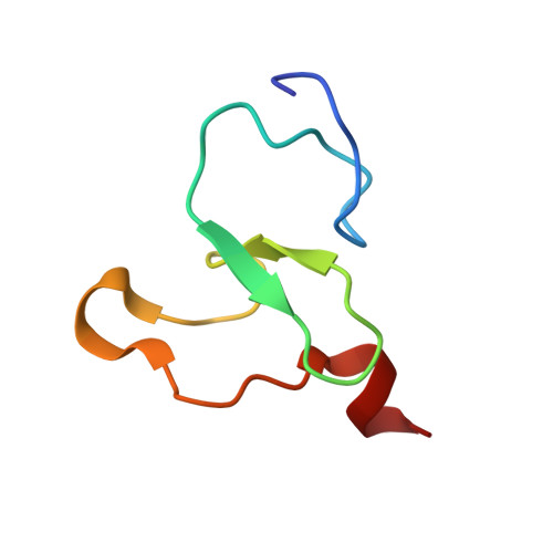
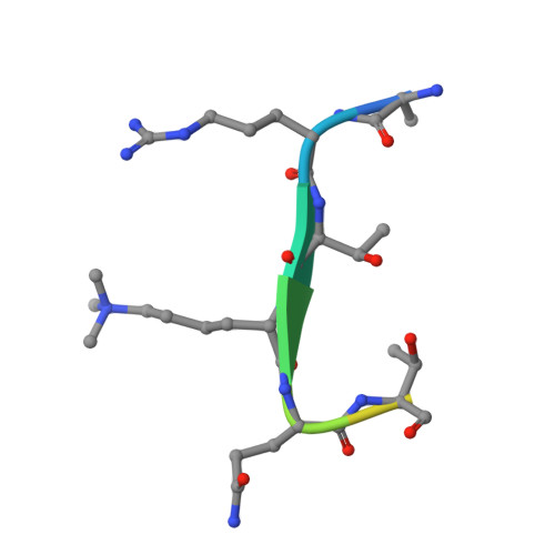
PHF13 is a molecular reader and transcriptional co-regulator of H3K4me2/3.

Chung, H.R.Xu, C.Fuchs, A.Mund, A.Lange, M.Staege, H.Schubert, T.Bian, C.Dunkel, I.Eberharter, A.Regnard, C.Klinker, H.Meierhofer, D.Cozzuto, L.Winterpacht, A.Di Croce, L.Min, J.Will, H.Kinkley, S.

(2016) Elife 5

  • DOI: https://doi.org/10.7554/eLife.10607
  • Primary Citation of Related Structures:  
    3O70, 3O7A

  • PubMed Abstract: 

    PHF13 is a chromatin affiliated protein with a functional role in differentiation, cell division, DNA damage response and higher chromatin order. To gain insight into PHF13's ability to modulate these processes, we elucidate the mechanisms targeting PHF13 to chromatin, its genome wide localization and its molecular chromatin context. Size exclusion chromatography, mass spectrometry, X-ray crystallography and ChIP sequencing demonstrate that PHF13 binds chromatin in a multivalent fashion via direct interactions with H3K4me2/3 and DNA, and indirectly via interactions with PRC2 and RNA PolII. Furthermore, PHF13 depletion disrupted the interactions between PRC2, RNA PolII S5P, H3K4me3 and H3K27me3 and resulted in the up and down regulation of genes functionally enriched in transcriptional regulation, DNA binding, cell cycle, differentiation and chromatin organization. Together our findings argue that PHF13 is an H3K4me2/3 molecular reader and transcriptional co-regulator, affording it the ability to impact different chromatin processes.


  • Organizational Affiliation

    Max Planck Institute for Molecular Genetics, Berlin, Germany.


Macromolecules
Find similar proteins by:  (by identity cutoff)  |  3D Structure
Entity ID: 1
MoleculeChains Sequence LengthOrganismDetailsImage
PHD finger protein 13 variant52Homo sapiensMutation(s): 0 
UniProt & NIH Common Fund Data Resources
Find proteins for Q86YI8 (Homo sapiens)
Explore Q86YI8 
Go to UniProtKB:  Q86YI8
PHAROS:  Q86YI8
GTEx:  ENSG00000116273 
Entity Groups  
Sequence Clusters30% Identity50% Identity70% Identity90% Identity95% Identity100% Identity
UniProt GroupQ86YI8
Sequence Annotations
Expand
  • Reference Sequence

Find similar proteins by:  Sequence   |   3D Structure  

Entity ID: 2
MoleculeChains Sequence LengthOrganismDetailsImage
H3K4ME3 HISTONE 11MER-PEPTIDE11Xenopus laevisMutation(s): 0 
UniProt
Find proteins for Q92133 (Xenopus laevis)
Explore Q92133 
Go to UniProtKB:  Q92133
Entity Groups  
Sequence Clusters30% Identity50% Identity70% Identity90% Identity95% Identity100% Identity
UniProt GroupQ92133
Sequence Annotations
Expand
  • Reference Sequence
Small Molecules
Modified Residues  1 Unique
IDChains TypeFormula2D DiagramParent
M3L
Query on M3L
B
L-PEPTIDE LINKINGC9 H21 N2 O2LYS
Experimental Data & Validation

Experimental Data

  • Method: X-RAY DIFFRACTION
  • Resolution: 1.67 Å
  • R-Value Free: 0.224 
  • R-Value Work: 0.183 
  • R-Value Observed: 0.185 
  • Space Group: P 21 21 21
Unit Cell:
Length ( Å )Angle ( ˚ )
a = 21.232α = 90
b = 44.334β = 90
c = 60.33γ = 90
Software Package:
Software NamePurpose
REFMACrefinement
PDB_EXTRACTdata extraction
HKL-2000data collection
HKL-2000data reduction
HKL-2000data scaling
MOLREPphasing

Structure Validation

View Full Validation Report


Entry History 

Revision History  (Full details and data files)

  • Version 1.0: 2010-10-06
    Type: Initial release
  • Version 1.1: 2011-07-13
    Changes: Version format compliance
  • Version 1.2: 2016-06-22
    Changes: Database references
  • Version 1.3: 2023-09-06
    Changes: Data collection, Database references, Derived calculations, Refinement description