3O0E

Crystal structure of OmpF in complex with colicin peptide OBS1


Experimental Data Snapshot

  • Method: X-RAY DIFFRACTION
  • Resolution: 3.01 Å
  • R-Value Free: 0.302 
  • R-Value Work: 0.265 
  • R-Value Observed: 0.267 

wwPDB Validation   3D Report Full Report


Ligand Structure Quality Assessment 


This is version 2.1 of the entry. See complete history


Literature

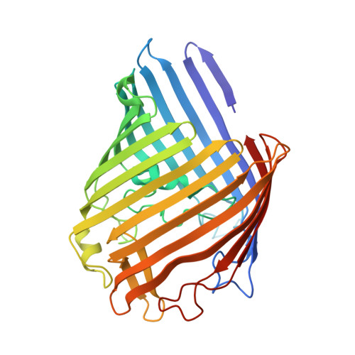
Directed epitope delivery across the Escherichia coli outer membrane through the porin OmpF.

Housden, N.G.Wojdyla, J.A.Korczynska, J.Grishkovskaya, I.Kirkpatrick, N.Brzozowski, A.M.Kleanthous, C.

(2010) Proc Natl Acad Sci U S A 107: 21412-21417

  • DOI: https://doi.org/10.1073/pnas.1010780107
  • Primary Citation of Related Structures:  
    3O0E

  • PubMed Abstract: 

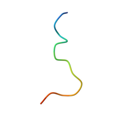
    The porins OmpF and OmpC are trimeric β-barrel proteins with narrow channels running through each monomer that exclude molecules > 600 Da while mediating the passive diffusion of small nutrients and metabolites across the Gram-negative outer membrane (OM). Here, we elucidate the mechanism by which an entire soluble protein domain (> 6 kDa) is delivered through the lumen of such porins. Following high-affinity binding to the vitamin B(12) receptor in Escherichia coli, the bacteriocin ColE9 recruits OmpF or OmpC using an 83-residue intrinsically unstructured translocation domain (IUTD) to deliver a 16-residue TolB-binding epitope (TBE) in the center of the IUTD to the periplasm where it triggers toxin entry. We demonstrate that the IUTD houses two OmpF-binding sites, OBS1 (residues 2-18) and OBS2 (residues 54-63), which flank the TBE and bind with K(d)s of 2 and 24 μM, respectively, at pH 6.5 and 25 ºC. We show the two OBSs share the same binding site on OmpF and that the colicin must house at least one of them for antibiotic activity. Finally, we report the structure of the OmpF-OBS1 complex that shows the colicin bound within the porin lumen spanning the membrane bilayer. Our study explains how colicins exploit porins to deliver epitope signals to the bacterial periplasm and, more broadly, how the inherent flexibility and narrow cross-sectional area of an IUP domain can endow it with the ability to traverse a biological membrane via the constricted lumen of a β-barrel membrane protein.


  • Organizational Affiliation

    Department of Biology, Area 10, and York Structural Biology Laboratory, University of York, Heslington, York YO10 5DD, United Kingdom.


Macromolecules
Find similar proteins by:  (by identity cutoff)  |  3D Structure
Entity ID: 1
MoleculeChains Sequence LengthOrganismDetailsImage
Porin OmpF
A, B, C, D, E
A, B, C, D, E, F
340Escherichia coliMutation(s): 0 
Membrane Entity: Yes 
UniProt
Find proteins for P02931 (Escherichia coli (strain K12))
Explore P02931 
Go to UniProtKB:  P02931
Entity Groups  
Sequence Clusters30% Identity50% Identity70% Identity90% Identity95% Identity100% Identity
UniProt GroupP02931
Sequence Annotations
Expand
  • Reference Sequence

Find similar proteins by:  Sequence   |   3D Structure  

Entity ID: 2
MoleculeChains Sequence LengthOrganismDetailsImage
Colicin-E917Escherichia coliMutation(s): 0 
EC: 3.1
UniProt
Find proteins for P09883 (Escherichia coli)
Explore P09883 
Go to UniProtKB:  P09883
Entity Groups  
Sequence Clusters30% Identity50% Identity70% Identity90% Identity95% Identity100% Identity
UniProt GroupP09883
Sequence Annotations
Expand
  • Reference Sequence
Experimental Data & Validation

Experimental Data

  • Method: X-RAY DIFFRACTION
  • Resolution: 3.01 Å
  • R-Value Free: 0.302 
  • R-Value Work: 0.265 
  • R-Value Observed: 0.267 
  • Space Group: P 1 21 1
Unit Cell:
Length ( Å )Angle ( ˚ )
a = 101.491α = 90
b = 101.618β = 94.46
c = 162.142γ = 90
Software Package:
Software NamePurpose
PHENIXrefinement
DENZOdata reduction
SCALEPACKdata scaling
PDB_EXTRACTdata extraction
ADSCdata collection
MOLREPphasing

Structure Validation

View Full Validation Report


Ligand Structure Quality Assessment 


Entry History & Funding Information

Deposition Data


Funding OrganizationLocationGrant Number
Wellcome TrustUnited Kingdom--
Biotechnology and Biological Sciences Research Council (BBSRC)United Kingdom--

Revision History  (Full details and data files)

  • Version 1.0: 2010-12-08
    Type: Initial release
  • Version 1.1: 2011-07-13
    Changes: Version format compliance
  • Version 1.2: 2020-07-29
    Type: Remediation
    Reason: Carbohydrate remediation
    Changes: Data collection, Derived calculations, Structure summary
  • Version 2.0: 2021-05-05
    Type: Coordinate replacement
    Reason: Model orientation/position
    Changes: Advisory, Atomic model, Author supporting evidence, Data collection, Database references, Derived calculations, Experimental preparation, Other, Refinement description, Source and taxonomy, Structure summary
  • Version 2.1: 2023-09-20
    Changes: Data collection, Database references, Refinement description