3NYX

Non-phosphorylated TYK2 JH1 domain with Quinoline-Thiadiazole-Thiophene Inhibitor


Experimental Data Snapshot

  • Method: X-RAY DIFFRACTION
  • Resolution: 2.50 Å
  • R-Value Free: 0.275 
  • R-Value Work: 0.215 
  • R-Value Observed: 0.219 

wwPDB Validation   3D Report Full Report


Ligand Structure Quality Assessment 


This is version 1.1 of the entry. See complete history


Literature

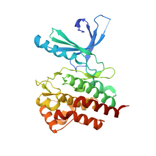
A new regulatory switch in a JAK protein kinase.

Tsui, V.Gibbons, P.Ultsch, M.Mortara, K.Chang, C.Blair, W.Pulk, R.Stanley, M.Starovasnik, M.Williams, D.Lamers, M.Leonard, P.Magnuson, S.Liang, J.Eigenbrot, C.

(2011) Proteins 79: 393-401

  • DOI: https://doi.org/10.1002/prot.22889
  • Primary Citation of Related Structures:  
    3NYX, 3NZ0

  • PubMed Abstract: 
  • Members of the JAK family of protein kinases mediate signal transduction from cytokine receptors to transcription factor activation. Over-stimulation of these pathways is causative in immune disorders like rheumatoid arthritis, psoriasis, lupus, and Crohn's disease ...

    Members of the JAK family of protein kinases mediate signal transduction from cytokine receptors to transcription factor activation. Over-stimulation of these pathways is causative in immune disorders like rheumatoid arthritis, psoriasis, lupus, and Crohn's disease. A search for selective inhibitors of a JAK kinase has led to our characterization of a previously unknown kinase conformation arising from presentation of Tyr962 of TYK2 to an inhibitory small molecule via an H-bonding interaction. A small minority of protein kinase domains has a Tyrosine residue in this position within the αC-β4 loop, and it is the only amino acid commonly seen here with H-bonding potential. These discoveries will aid design of inhibitors that discriminate among the JAK family and more widely among protein kinases.


    Organizational Affiliation

    Department of Discovery Chemistry, Genentech, Inc, South San Francisco, California 94080, USA.


Macromolecules
Find similar proteins by:  (by identity cutoff)  |  3D Structure
Entity ID: 1
MoleculeChainsSequence LengthOrganismDetailsImage
Non-receptor tyrosine-protein kinase TYK2302Homo sapiensMutation(s): 1 
Gene Names: TYK2
EC: 2.7.10.2
UniProt & NIH Common Fund Data Resources
Find proteins for P29597 (Homo sapiens)
Explore P29597 
Go to UniProtKB:  P29597
PHAROS:  P29597
Entity Groups  
Sequence Clusters30% Identity50% Identity70% Identity90% Identity95% Identity100% Identity
UniProt GroupP29597
Protein Feature View
Expand
  • Reference Sequence
Small Molecules
Ligands 2 Unique
IDChainsName / Formula / InChI Key2D Diagram3D Interactions
TZ1
Query on TZ1

Download Ideal Coordinates CCD File 
B [auth A]N-{5-[(7-chloroquinolin-4-yl)sulfanyl]-1,3,4-thiadiazol-2-yl}thiophene-2-carboxamide
C16 H9 Cl N4 O S3
KWDHWMGDHBBJIA-UHFFFAOYSA-N
 Ligand Interaction
DTT
Query on DTT

Download Ideal Coordinates CCD File 
C [auth A],
D [auth A]
2,3-DIHYDROXY-1,4-DITHIOBUTANE
C4 H10 O2 S2
VHJLVAABSRFDPM-IMJSIDKUSA-N
 Ligand Interaction
Binding Affinity Annotations 
IDSourceBinding Affinity
TZ1 PDBBind:  3NYX Ki: 32 (nM) from 1 assay(s)
Binding MOAD:  3NYX Ki: 32 (nM) from 1 assay(s)
Experimental Data & Validation

Experimental Data

  • Method: X-RAY DIFFRACTION
  • Resolution: 2.50 Å
  • R-Value Free: 0.275 
  • R-Value Work: 0.215 
  • R-Value Observed: 0.219 
  • Space Group: P 21 21 21
Unit Cell:
Length ( Å )Angle ( ˚ )
a = 55.318α = 90
b = 70.436β = 90
c = 86.488γ = 90
Software Package:
Software NamePurpose
BOSdata collection
PHASERphasing
REFMACrefinement
HKL-2000data reduction
SCALEPACKdata scaling

Structure Validation

View Full Validation Report


Ligand Structure Quality Assessment 


Entry History 

Deposition Data

Revision History  (Full details and data files)

  • Version 1.0: 2010-10-20
    Type: Initial release
  • Version 1.1: 2011-07-13
    Changes: Version format compliance