


Aminoindazole PDK1 Inhibitors: A Case Study in Fragment-Based Drug Discovery
Medina, J.R., Blackledge, C.W., Heerding, D.A., Campobasso, N., Ward, P., Briand, J., Wright, L., Axten, J.M.To be published.
Experimental Data Snapshot
Entity ID: 1 | |||||
|---|---|---|---|---|---|
| Molecule | Chains | Sequence Length | Organism | Details | Image |
| PkB-like | 292 | Homo sapiens | Mutation(s): 0 Gene Names: PkB-like 1 EC: 2.7.11.1 | 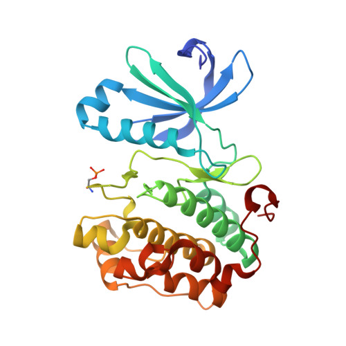 | |
UniProt & NIH Common Fund Data Resources | |||||
PHAROS: O15530 GTEx: ENSG00000140992 | |||||
Entity Groups | |||||
| Sequence Clusters | 30% Identity50% Identity70% Identity90% Identity95% Identity100% Identity | ||||
| UniProt Group | O15530 | ||||
Sequence AnnotationsExpand | |||||
Reference Sequence | |||||
| Ligands 3 Unique | |||||
|---|---|---|---|---|---|
| ID | Chains | Name / Formula / InChI Key | 2D Diagram | 3D Interactions | |
| JMZ Download:Ideal Coordinates CCD File | L [auth A] | 6-(2-aminopyrimidin-4-yl)-1H-indazol-3-amine C11 H10 N6 QVWIVWPIJUYRRF-UHFFFAOYSA-N |  | ||
| SO4 Download:Ideal Coordinates CCD File | F [auth A] G [auth A] H [auth A] I [auth A] J [auth A] | SULFATE ION O4 S QAOWNCQODCNURD-UHFFFAOYSA-L |  | ||
| GOL Download:Ideal Coordinates CCD File | B [auth A], C [auth A], D [auth A], E [auth A] | GLYCEROL C3 H8 O3 PEDCQBHIVMGVHV-UHFFFAOYSA-N |  | ||
| Modified Residues 1 Unique | |||||
|---|---|---|---|---|---|
| ID | Chains | Type | Formula | 2D Diagram | Parent |
| SEP Query on SEP | A | L-PEPTIDE LINKING | C3 H8 N O6 P |  | SER |
| Length ( Å ) | Angle ( ˚ ) |
|---|---|
| a = 122.761 | α = 90 |
| b = 122.761 | β = 90 |
| c = 47.106 | γ = 120 |
| Software Name | Purpose |
|---|---|
| DENZO | data reduction |
| SCALEPACK | data scaling |
| PHENIX | refinement |
| PDB_EXTRACT | data extraction |
| ADSC | data collection |
| PHASER | phasing |


