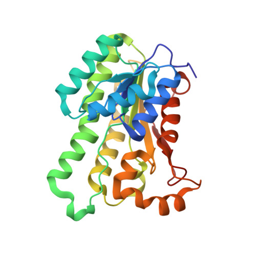
3NUG

Crystal structure of wild type tetrameric pyridoxal 4-dehydrogenase from Mesorhizobium loti


Experimental Data Snapshot

  • Method: X-RAY DIFFRACTION
  • Resolution: 1.79 Å
  • R-Value Free: 0.202 
  • R-Value Work: 0.152 
  • R-Value Observed: 0.155 

wwPDB Validation   3D Report Full Report


Ligand Structure Quality Assessment 


This is version 1.3 of the entry. See complete history


Literature

The crystal structure of SDR-type pyridoxal 4-dehydrogenase of Mesorhizobium loti

Chu, H.N.Kobayashi, J.Mikami, B.Yagi, T.

(2011) Biosci Biotechnol Biochem 75: 388-390

  • DOI: https://doi.org/10.1271/bbb.100748
  • Primary Citation of Related Structures:  
    3NUG

  • PubMed Abstract: 

    Pyridoxal 4-dehydrogenase catalyzes the irreversible oxidation of pyridoxal to 4-pyridoxolactone and is involved in degradation pathway I of pyridoxine, a vitamin B(6) compound. Its crystal structure was elucidated for the first time. Molecular replacement with (S)-1-phenylthanol dehydrogenase (PDB code 2EW8) was adopted to determine the tertiary structure of the NAD(+)-bound enzyme.


  • Organizational Affiliation

    Faculty of Agriculture and Agricultural Science Program, Graduate School of Integral Arts and Science, Kochi University, Nankoku, Kochi, Japan.


Macromolecules
Find similar proteins by:  (by identity cutoff)  |  3D Structure
Entity ID: 1
MoleculeChains Sequence LengthOrganismDetailsImage
3-oxoacyl-(Acyl-carrier protein) reductase
A, B, C, D
247Mesorhizobium lotiMutation(s): 0 
Gene Names: mlr6807pldh-t
EC: 1.1.1.107
UniProt
Find proteins for Q988B7 (Mesorhizobium japonicum (strain LMG 29417 / CECT 9101 / MAFF 303099))
Explore Q988B7 
Go to UniProtKB:  Q988B7
Entity Groups  
Sequence Clusters30% Identity50% Identity70% Identity90% Identity95% Identity100% Identity
UniProt GroupQ988B7
Sequence Annotations
Expand
  • Reference Sequence
Experimental Data & Validation

Experimental Data

  • Method: X-RAY DIFFRACTION
  • Resolution: 1.79 Å
  • R-Value Free: 0.202 
  • R-Value Work: 0.152 
  • R-Value Observed: 0.155 
  • Space Group: P 1 21 1
Unit Cell:
Length ( Å )Angle ( ˚ )
a = 85.638α = 90
b = 50.695β = 89.29
c = 94.551γ = 90
Software Package:
Software NamePurpose
HKL-2000data collection
PHASERphasing
PHENIXrefinement
HKL-2000data reduction
HKL-2000data scaling

Structure Validation

View Full Validation Report


Ligand Structure Quality Assessment 


Entry History 

Deposition Data

  • Released Date: 2011-04-20 
  • Deposition Author(s): Chu, H.N.

Revision History  (Full details and data files)

  • Version 1.0: 2011-04-20
    Type: Initial release
  • Version 1.1: 2011-07-13
    Changes: Version format compliance
  • Version 1.2: 2017-11-08
    Changes: Advisory, Refinement description
  • Version 1.3: 2023-11-01
    Changes: Advisory, Data collection, Database references, Derived calculations, Refinement description