3NQ1 | pdb_00003nq1

Crystal Structure of Tyrosinase from Bacillus megaterium in complex with inhibitor kojic acid


Experimental Data Snapshot

  • Method: X-RAY DIFFRACTION
  • Resolution: 2.30 Å
  • R-Value Free: 
    0.294 (Depositor), 0.294 (DCC) 
  • R-Value Work: 
    0.260 (Depositor), 0.264 (DCC) 
  • R-Value Observed: 
    0.262 (Depositor) 

wwPDB Validation 3D Report Full Report

Validation slider image for 3NQ1

This is version 1.3 of the entry. See complete history

Literature

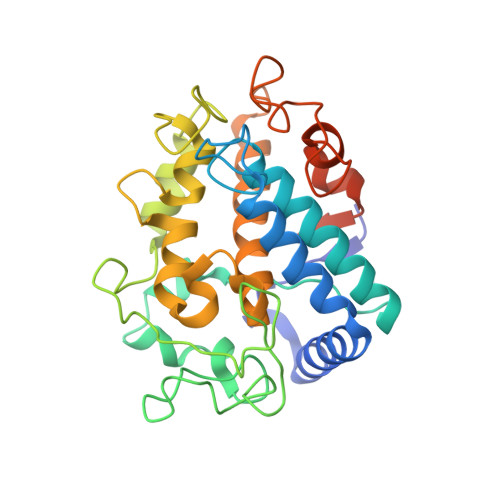
First structures of an active bacterial tyrosinase reveal copper plasticity.

Sendovski, M.Kanteev, M.Shuster Ben-Yosef, V.Adir, N.Fishman, A.

(2011) J Mol Biology 405: 227-237

  • DOI: https://doi.org/10.1016/j.jmb.2010.10.048
  • Primary Citation Related Structures: 
    3NM8, 3NPY, 3NQ0, 3NQ1, 3NQ5, 3NTM

  • PubMed Abstract: 

    Tyrosinase is a member of the type 3 copper enzyme family that is involved in the production of melanin in a wide range of organisms. The crystal structures of a tyrosinase from Bacillus megaterium were determined at a resolution of 2.0-2.3 Å. The enzyme crystallized as a dimer in the asymmetric unit and was shown to be active in crystal. The overall monomeric structure is similar to that of the monomer of the previously determined tyrosinase from Streptomyces castaneoglobisporus, but it does not contain an accessory Cu-binding "caddie" protein. Two Cu(II) ions, serving as the major cofactors within the active site, are coordinated by six conserved histidine residues. However, determination of structures under different conditions shows varying occupancies and positions of the copper ions. This apparent mobility in copper binding modes indicates that there is a pathway by which copper is accumulated or lost by the enzyme. Additionally, we suggest that residues R209 and V218, situated in a second shell of residues surrounding the active site, play a role in substrate binding orientation based on their flexibility and position. The determination of a structure with the inhibitor kojic acid, the first tyrosinase structure with a bound ligand, revealed additional residues involved in the positioning of substrates in the active site. Comparison of wild-type structures with the structure of the site-specific variant R209H, which possesses a higher monophenolase/diphenolase activity ratio, lends further support to a previously suggested mechanism by which monophenolic substrates dock mainly to CuA.


  • Organizational Affiliation
    • Department of Biotechnology and Food Engineering, Technion-Israel Institute of Technology, Haifa 32000, Israel.

Macromolecule Content 

  • Total Structure Weight: 71.96 kDa 
  • Atom Count: 4,885 
  • Modeled Residue Count: 566 
  • Deposited Residue Count: 606 
  • Unique protein chains: 1

Macromolecules

Find similar proteins by:|  3D Structure
Entity ID: 1
MoleculeChains  Sequence LengthOrganismDetailsImage
Tyrosinase
A, B
303Priestia megateriumMutation(s): 0 
EC: 1.14.18.1
UniProt
Find proteins for B2ZB02 (Priestia megaterium)
Explore B2ZB02 
Go to UniProtKB:  B2ZB02
Entity Groups
Sequence Clusters30% Identity50% Identity70% Identity90% Identity95% Identity100% Identity
UniProt GroupB2ZB02
Sequence Annotations
Expand
Reference Sequence

Small Molecules

Ligands 3 Unique
IDChains Name / Formula / InChI Key2D Diagram3D Interactions
KOJ

Query on KOJ


Download:Ideal Coordinates CCD File
F [auth A],
Q [auth B]
5-HYDROXY-2-(HYDROXYMETHYL)-4H-PYRAN-4-ONE
C6 H6 O4
BEJNERDRQOWKJM-UHFFFAOYSA-N
ZN

Query on ZN


Download:Ideal Coordinates CCD File
E [auth A]
G [auth A]
H [auth A]
I [auth A]
J [auth A]
E [auth A],
G [auth A],
H [auth A],
I [auth A],
J [auth A],
K [auth A],
L [auth A],
M [auth A],
P [auth B],
R [auth B],
S [auth B],
T [auth B],
U [auth B]
ZINC ION
Zn
PTFCDOFLOPIGGS-UHFFFAOYSA-N
CU

Query on CU


Download:Ideal Coordinates CCD File
C [auth A],
D [auth A],
N [auth B],
O [auth B]
COPPER (II) ION
Cu
JPVYNHNXODAKFH-UHFFFAOYSA-N
Binding Affinity Annotations 
IDSourceBinding Affinity
KOJ BindingDB:  3NQ1 Kd: 3.77e+5 (nM) from 1 assay(s)
IC50: 5.20e+4 (nM) from 1 assay(s)

Experimental Data & Validation

Experimental Data

  • Method: X-RAY DIFFRACTION
  • Resolution: 2.30 Å
  • R-Value Free:  0.294 (Depositor), 0.294 (DCC) 
  • R-Value Work:  0.260 (Depositor), 0.264 (DCC) 
  • R-Value Observed: 0.262 (Depositor) 
Space Group: P 21 21 21
Unit Cell:
Length ( Å )Angle ( ˚ )
a = 51.38α = 90
b = 83.53β = 90
c = 146.16γ = 90
Software Package:
Software NamePurpose
MOSFLMdata reduction
SCALAdata scaling
PHASERphasing
REFMACrefinement
PDB_EXTRACTdata extraction
DNAdata collection
PHENIXrefinement

Structure Validation

View Full Validation Report


Entry History 

Deposition Data

Revision History  (Full details and data files)

  • Version 1.0: 2010-11-17
    Type: Initial release
  • Version 1.1: 2011-07-13
    Changes: Version format compliance
  • Version 1.2: 2014-02-26
    Changes: Database references
  • Version 1.3: 2023-12-27
    Changes: Data collection, Database references, Derived calculations