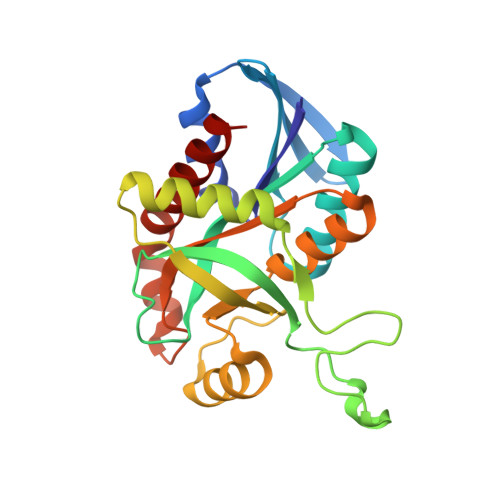
Enzyme-ligand interactions that drive active site rearrangements in the Helicobacter pylori 5'-methylthioadenosine/S-adenosylhomocysteine nucleosidase.
Ronning, D.R., Iacopelli, N.M., Mishra, V.(2010) Protein Sci 19: 2498-2510
- PubMed: 20954236 Search on PubMedSearch on PubMed Central
- DOI: https://doi.org/10.1002/pro.524
- Primary Citation Related Structures:
3NM4, 3NM5, 3NM6 - PubMed Abstract:
The bacterial enzyme 5'-methylthioadenosine/S-adenosylhomocysteine nucleosidase (MTAN) plays a central role in three essential metabolic pathways in bacteria: methionine salvage, purine salvage, and polyamine biosynthesis. Recently, its role in the pathway that leads to the production of autoinducer II, an important component in quorum-sensing, has garnered much interest. Because of this variety of roles, MTAN is an attractive target for developing new classes of inhibitors that influence bacterial virulence and biofilm formation. To gain insight toward the development of new classes of MTAN inhibitors, the interactions between the Helicobacter pylori-encoded MTAN and its substrates and substrate analogs were probed using X-ray crystallography. The structures of MTAN, an MTAN-Formycin A complex, and an adenine bound form were solved by molecular replacement and refined to 1.7, 1.8, and 1.6 Å, respectively. The ribose-binding site in the MTAN and MTAN-adenine cocrystal structures contain a tris[hydroxymethyl]aminomethane molecule that stabilizes the closed form of the enzyme and displaces a nucleophilic water molecule necessary for catalysis. This research gives insight to the interactions between MTAN and bound ligands that promote closing of the enzyme active site and highlights the potential for designing new classes of MTAN inhibitors using a link/grow or ligand assembly development strategy based on the described H. pylori MTAN crystal structures.
- Department of Chemistry, University of Toledo, Toledo, Ohio 43606, USA. donald.ronning@utoledo.edu
Organizational Affiliation: