


Crystal structure of the VP0956 protein from Vibrio parahaemolyticus.
Vorobiev, S., Neely, H., Seetharaman, J., Wang, D., Mao, L., Xiao, R., Acton, T.B., Montelione, G.T., Tong, L., Hunt, J.To be published.
Experimental Data Snapshot
Entity ID: 1 | |||||
|---|---|---|---|---|---|
| Molecule | Chains | Sequence Length | Organism | Details | Image |
| Uncharacterized protein VP0956 | 549 | Vibrio parahaemolyticus | Mutation(s): 1 Gene Names: VP0956 | 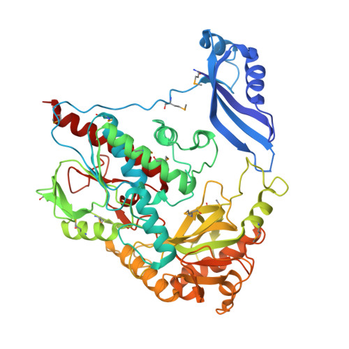 | |
UniProt | |||||
Entity Groups | |||||
| Sequence Clusters | 30% Identity50% Identity70% Identity90% Identity95% Identity100% Identity | ||||
| UniProt Group | Q87R42 | ||||
Sequence AnnotationsExpand | |||||
Reference Sequence | |||||
| Ligands 1 Unique | |||||
|---|---|---|---|---|---|
| ID | Chains | Name / Formula / InChI Key | 2D Diagram | 3D Interactions | |
| FAD Download:Ideal Coordinates CCD File | B [auth A] | FLAVIN-ADENINE DINUCLEOTIDE C27 H33 N9 O15 P2 VWWQXMAJTJZDQX-UYBVJOGSSA-N |  | ||
| Modified Residues 1 Unique | |||||
|---|---|---|---|---|---|
| ID | Chains | Type | Formula | 2D Diagram | Parent |
| MSE Query on MSE | A | L-PEPTIDE LINKING | C5 H11 N O2 Se |  | MET |
| Length ( Å ) | Angle ( ˚ ) |
|---|---|
| a = 69.079 | α = 90 |
| b = 78.166 | β = 90 |
| c = 101.875 | γ = 90 |
| Software Name | Purpose |
|---|---|
| ADSC | data collection |
| SHELXDE | phasing |
| PHENIX | refinement |
| DENZO | data reduction |
| SCALEPACK | data scaling |


