3NFQ | pdb_00003nfq

Crystal structure of the conserved central domain of yeast Spn1/Iws1


Experimental Data Snapshot

  • Method: X-RAY DIFFRACTION
  • Resolution: 1.85 Å
  • R-Value Free: 
    0.265 (Depositor), 0.264 (DCC) 
  • R-Value Work: 
    0.203 (Depositor), 0.203 (DCC) 
  • R-Value Observed: 
    0.207 (Depositor) 

wwPDB Validation 3D Report Full Report

Validation slider image for 3NFQ

This is version 1.2 of the entry. See complete history

Literature

The Transcription Factor Spn1 Regulates Gene Expression via a Highly Conserved Novel Structural Motif.

Pujari, V.Radebaugh, C.A.Chodaparambil, J.V.Muthurajan, U.M.Almeida, A.R.Fischbeck, J.A.Luger, K.Stargell, L.A.

(2010) J Mol Biology 404: 1-15

  • DOI: https://doi.org/10.1016/j.jmb.2010.09.040
  • Primary Citation Related Structures: 
    3NFQ

  • PubMed Abstract: 

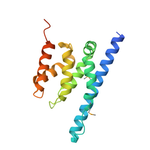
    Spn1/Iws1 plays essential roles in the regulation of gene expression by RNA polymerase II (RNAPII), and it is highly conserved in organisms ranging from yeast to humans. Spn1 physically and/or genetically interacts with RNAPII, TBP (TATA-binding protein), TFIIS (transcription factor IIS), and a number of chromatin remodeling factors (Swi/Snf and Spt6). The central domain of Spn1 (residues 141-305 out of 410) is necessary and sufficient for performing the essential functions of SPN1 in yeast cells. Here, we report the high-resolution (1.85 Å) crystal structure of the conserved central domain of Saccharomyces cerevisiae Spn1. The central domain is composed of eight α-helices in a right-handed superhelical arrangement and exhibits structural similarity to domain I of TFIIS. A unique structural feature of Spn1 is a highly conserved loop, which defines one side of a pronounced cavity. The loop and the other residues forming the cavity are highly conserved at the amino acid level among all Spn1 family members, suggesting that this is a signature motif for Spn1 orthologs. The locations and the molecular characterization of temperature-sensitive mutations in Spn1 indicate that the cavity is a key attribute of Spn1 that is critical for its regulatory functions during RNAPII-mediated transcriptional activity.


  • Organizational Affiliation
    • Department of Biochemistry and Molecular Biology, Colorado State University, Fort Collins, CO 80523-1870, USA.

Macromolecule Content 

  • Total Structure Weight: 39.37 kDa 
  • Atom Count: 2,718 
  • Modeled Residue Count: 290 
  • Deposited Residue Count: 340 
  • Unique protein chains: 1

Macromolecules

Find similar proteins by:|  3D Structure
Entity ID: 1
MoleculeChains  Sequence LengthOrganismDetailsImage
Transcription factor IWS1
A, B
170Saccharomyces cerevisiaeMutation(s): 0 
Gene Names: IWS1SPN1YPR133C
UniProt
Find proteins for Q06505 (Saccharomyces cerevisiae (strain ATCC 204508 / S288c))
Explore Q06505 
Go to UniProtKB:  Q06505
Entity Groups
Sequence Clusters30% Identity50% Identity70% Identity90% Identity95% Identity100% Identity
UniProt GroupQ06505
Sequence Annotations
Expand
Reference Sequence

Small Molecules

Modified Residues  1 Unique
IDChains TypeFormula2D DiagramParent
MSE
Query on MSE
A, B
L-PEPTIDE LINKINGC5 H11 N O2 SeMET

Experimental Data & Validation

Experimental Data

  • Method: X-RAY DIFFRACTION
  • Resolution: 1.85 Å
  • R-Value Free:  0.265 (Depositor), 0.264 (DCC) 
  • R-Value Work:  0.203 (Depositor), 0.203 (DCC) 
  • R-Value Observed: 0.207 (Depositor) 
Space Group: P 21 21 21
Unit Cell:
Length ( Å )Angle ( ˚ )
a = 63.6α = 90
b = 67.8β = 90
c = 76γ = 90
Software Package:
Software NamePurpose
CrystalCleardata collection
SOLVEphasing
REFMACrefinement
d*TREKdata reduction
d*TREKdata scaling

Structure Validation

View Full Validation Report


Entry History 

Deposition Data

Revision History  (Full details and data files)

  • Version 1.0: 2010-10-13
    Type: Initial release
  • Version 1.1: 2011-07-13
    Changes: Version format compliance
  • Version 1.2: 2024-10-30
    Changes: Data collection, Database references, Derived calculations, Structure summary