Comparison of Cyclooxygenase-1 Crystal Structures: Cross-Talk between Monomers Comprising Cyclooxygenase-1 Homodimers
Sidhu, R.S., Lee, J.Y., Yuan, C., Smith, W.L.(2010) Biochemistry 49: 7069-7079
- PubMed: 20669977 Search on PubMedSearch on PubMed Central
- DOI: https://doi.org/10.1021/bi1003298
- Primary Citation Related Structures:
3N8V, 3N8W, 3N8X, 3N8Y, 3N8Z - PubMed Abstract:
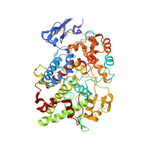
Prostaglandin endoperoxide H synthases (PGHSs)-1 and -2 (also called cyclooxygenases (COXs)-1 and -2) catalyze the committed step in prostaglandin biosynthesis. Both isoforms are targets of nonsteroidal antiinflammatory drugs (NSAIDs). PGHSs are homodimers that exhibit half-of-sites COX activity; moreover, some NSAIDs cause enzyme inhibition by binding only one monomer. To learn more about the cross-talk that must be occurring between the monomers comprising each PGHS-1 dimer, we analyzed structures of PGHS-1 crystallized under five different conditions including in the absence of any tightly binding ligand and in the presence of nonspecific NSAIDs and of a COX-2 inhibitor. When crystallized with substoichiometric amounts of an NSAID, both monomers are often fully occupied with inhibitor; thus, the enzyme prefers to crystallize in a fully occupied form. In comparing the five structures, we only observe changes in the positions of residues 123-129 and residues 510-515. In cases where one monomer is fully occupied with an NSAID and the partner monomer is incompletely occupied, an alternate conformation of the loop involving residues 123-129 is seen in the partially occupied monomer. We propose, on the basis of this observation and previous cross-linking studies, that cross-talk between monomers involves this mobile 123-129 loop, which is located at the dimer interface. In ovine PGHS-1 crystallized in the absence of an NSAID, there is an alternative route for substrate entry into the COX site different than the well-known route through the membrane binding domain.
- Department of Biological Chemistry, University of Michigan, Ann Arbor, Michigan 48109, USA.
Organizational Affiliation: