3N76

Crystal structure of 3-dehydroquinate dehydratase from Mycobacterium tuberculosis in complex with compound 5


Experimental Data Snapshot

  • Method: X-RAY DIFFRACTION
  • Resolution: 1.90 Å
  • R-Value Free: 0.221 
  • R-Value Work: 0.175 
  • R-Value Observed: 0.178 

wwPDB Validation   3D Report Full Report


Ligand Structure Quality Assessment 


This is version 1.4 of the entry. See complete history


Literature

Structural investigation of inhibitor designs targeting 3-dehydroquinate dehydratase from the shikimate pathway of Mycobacterium tuberculosis.

Dias, M.V.Snee, W.C.Bromfield, K.M.Payne, R.J.Palaninathan, S.K.Ciulli, A.Howard, N.I.Abell, C.Sacchettini, J.C.Blundell, T.L.

(2011) Biochem J 436: 729-739

  • DOI: https://doi.org/10.1042/BJ20110002
  • Primary Citation of Related Structures:  
    3N59, 3N76, 3N7A, 3N86, 3N87, 3N8K, 3N8N

  • PubMed Abstract: 

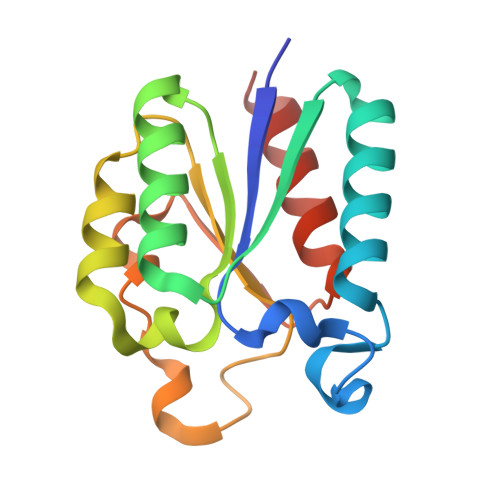
    The shikimate pathway is essential in Mycobacterium tuberculosis and its absence from humans makes the enzymes of this pathway potential drug targets. In the present paper, we provide structural insights into ligand and inhibitor binding to 3-dehydroquinate dehydratase (dehydroquinase) from M. tuberculosis (MtDHQase), the third enzyme of the shikimate pathway. The enzyme has been crystallized in complex with its reaction product, 3-dehydroshikimate, and with six different competitive inhibitors. The inhibitor 2,3-anhydroquinate mimics the flattened enol/enolate reaction intermediate and serves as an anchor molecule for four of the inhibitors investigated. MtDHQase also forms a complex with citrazinic acid, a planar analogue of the reaction product. The structure of MtDHQase in complex with a 2,3-anhydroquinate moiety attached to a biaryl group shows that this group extends to an active-site subpocket inducing significant structural rearrangement. The flexible extensions of inhibitors designed to form π-stacking interactions with the catalytic Tyr24 have been investigated. The high-resolution crystal structures of the MtDHQase complexes provide structural evidence for the role of the loop residues 19-24 in MtDHQase ligand binding and catalytic mechanism and provide a rationale for the design and efficacy of inhibitors.


  • Organizational Affiliation

    Department of Biochemistry, University of Cambridge, 80 Tennis Court Road, Cambridge CB2 1GA, UK.


Macromolecules
Find similar proteins by:  (by identity cutoff)  |  3D Structure
Entity ID: 1
MoleculeChains Sequence LengthOrganismDetailsImage
3-dehydroquinate dehydratase147Mycobacterium tuberculosisMutation(s): 0 
Gene Names: aroDaroQMT2612MTCY159.19Rv2537c
EC: 4.2.1.10
UniProt
Find proteins for P9WPX7 (Mycobacterium tuberculosis (strain ATCC 25618 / H37Rv))
Explore P9WPX7 
Go to UniProtKB:  P9WPX7
Entity Groups  
Sequence Clusters30% Identity50% Identity70% Identity90% Identity95% Identity100% Identity
UniProt GroupP9WPX7
Sequence Annotations
Expand
  • Reference Sequence
Small Molecules
Ligands 1 Unique
IDChains Name / Formula / InChI Key2D Diagram3D Interactions
CA2
Query on CA2

Download Ideal Coordinates CCD File 
B [auth A](1S,3R,4R,5S)-1,3,4-TRIHYDROXY-5-(3-PHENOXYPROPYL)CYCLOHEXANECARBOXYLIC ACID
C16 H22 O6
SCUFESRLGCQXRX-DCDXPUDHSA-N
Binding Affinity Annotations 
IDSourceBinding Affinity
CA2 PDBBind:  3N76 Ki: 140 (nM) from 1 assay(s)
Binding MOAD:  3N76 Ki: 140 (nM) from 1 assay(s)
Experimental Data & Validation

Experimental Data

  • Method: X-RAY DIFFRACTION
  • Resolution: 1.90 Å
  • R-Value Free: 0.221 
  • R-Value Work: 0.175 
  • R-Value Observed: 0.178 
  • Space Group: F 2 3
Unit Cell:
Length ( Å )Angle ( ˚ )
a = 126.41α = 90
b = 126.41β = 90
c = 126.41γ = 90
Software Package:
Software NamePurpose
HKL-2000data collection
AMoREphasing
REFMACrefinement
MOSFLMdata reduction
SCALAdata scaling

Structure Validation

View Full Validation Report


Ligand Structure Quality Assessment 


Entry History 

Deposition Data

Revision History  (Full details and data files)

  • Version 1.0: 2011-05-11
    Type: Initial release
  • Version 1.1: 2011-07-13
    Changes: Version format compliance
  • Version 1.2: 2012-02-29
    Changes: Structure summary
  • Version 1.3: 2018-01-24
    Changes: Structure summary
  • Version 1.4: 2023-09-06
    Changes: Data collection, Database references, Derived calculations, Refinement description